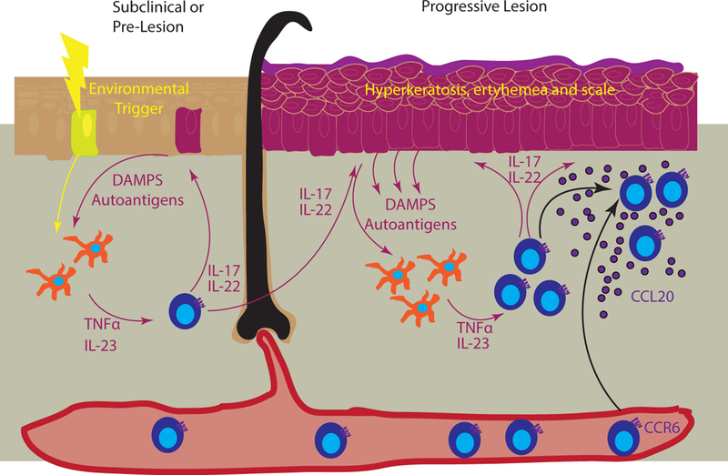
Figure 4. Chemokines in developing psoriatic lesions.

Left panel represents the precipitation of psoriasis in subclinical skin, where an environmental trigger (yellow bolt) induces release of danger associated molecular patterns (DAMPs) and autoantigens that are sensed by dermal APCs (yellow cells). These in turn secrete TNFα and IL-23, which activate skin T cells to produce IL-17 and IL-22, creating a feedforward loop (represented by magenta arrows). These cytokines in turn continue to damage keratinocytes, which results in the release of additional DAMPs and potential autoantigens. Right panel depicts a progressive lesion, with hyperkeratosis, erythema, and scale (magenta epidermis). T cell recruitment to the skin is primarily through keratinocyte-derived CCL20 (purple) that recruits CCR6-bearing cells.
