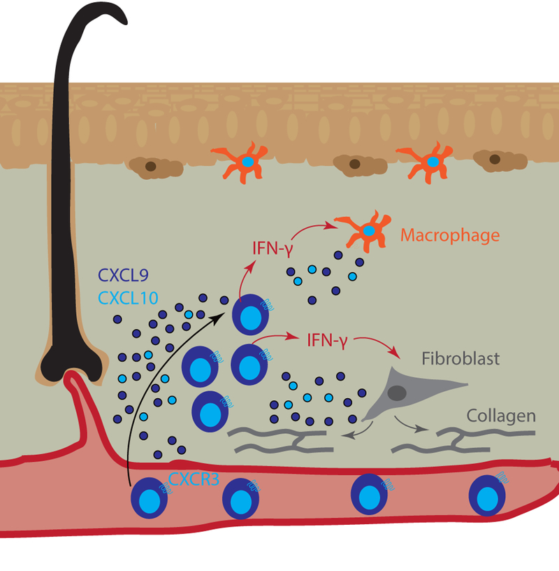
Figure 5. Chemokines promote T cell recruitment and fibrotic inflammation in morphea.

Morphea is characterized by deep dermal infiltrates and fibrosis, which are comprised primarily of T cells and macrophages (orange dermal APC) that may act directly upon fibroblasts (grey cells). Reports indicate that CXCR3 ligands are highly induced within lesional skin and serve as biomarkers of disease. These ligands recruit T cells (blue), which secrete IFN-γ (red arrows) to activate dermal macrophages (orange cells). Macrophages secrete additional CXCR3 ligands (CXCL9 dark blue, CXCL10 light blue dots) to further recruit T cells. Fibroblasts become activated by signals from profibrotic T cells and produce collagen (grey lines/arrows) to form fibrotic plaques.
