
Histologic features of human NASH.
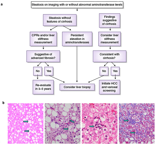
a, A schema for diagnosing NAFLD and NASH. Credit: Marina Corral Spence/Springer Nature. b, The panel of images from liver biopsies demonstrate the typical appearances of macrovesicular steatosis (fat), hepatocellular ballooning, lobular inflammation and pericellular fibrosis (arrows). As the disease progresses into cirrhosis (not shown), these features may also regress. H&E staining images were courtesy of Pierre Bedossa)