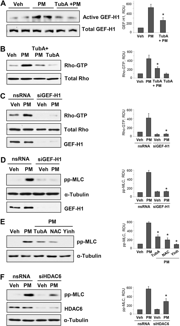
Figure 4. Inhibition of HDAC6 attenuates PM-induced MT destabilization and Rho activation.
Cells were pretreated with 10 μM of TubA for 30 min followed by PM stimulation (20 μg/cm2, 15 min). A - GEF-H1 pulldown assay was performed to capture active GEF-H1 with immobilized RhoG17A protein beads. B - Rho-GTP pulldown assay was performed using Rhotekin beads. Total GEF-H1 and Rho proteins in respective groups were used as normalization controls. C - Rho-GTP pulldown assay of pulmonary EC transfected with nonspecific (nsRNA) or GEF-H1-specific siRNA (siGEF-H1) and stimulated with PM (20 μg/cm2, 15 min). D - Western blot analysis of phospho-MLC levels. Cells were treated as above. Efficiency of GEF-H1 knockdown was confirmed by western blot with GEF-Ha antibody. Western blot analysis of RhoA in total cell lysates and membrane reprobing for α-tubulin was used as normalization controls. E - Cells were pretreated with HDAC6 inhibitor TubA (10 μM), Rho kinase inhibitor Y27632 (2 μM) or ROS inhibitor NAC (1 mM) for 30 min followed by PM challenge (20 μg/cm2, 6 hrs) and analysis of MLC phosphorylation by Western blot. F - Cells were transfected with non-specific or siRNA specific to HDAC6 (siHDAC6) followed by stimulation with PM (20 μg/cm2, 6 hrs). Phospho-MLC levels were detected in cell lysates by Western blot with phospho-MLC antibody. Membrane reprobing with α-tubulin and HDAC6 antibodies was performed to verify equal loading and HDAC6 knockdown, respectively. Corresponding bar graphs depict quantitative densitometry analysis of western blot experiments; n=3; **p<0.05. Data are expressed as mean + SD.

