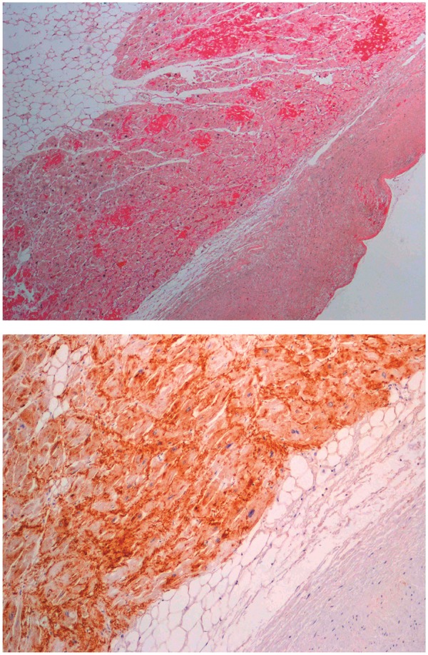
Figure 2.

Sirius red staining of left atrial tissue showing high grade isolated atrial amyloidosis (x4) (upper panel). Immunostaining of left atrial tissue in a patient with isolated atrial amyloidosis demonstrating the presence of atrial natriuretic peptide (x4) (lower panel). Reprinted with permission of Ariyarajah et al.23
