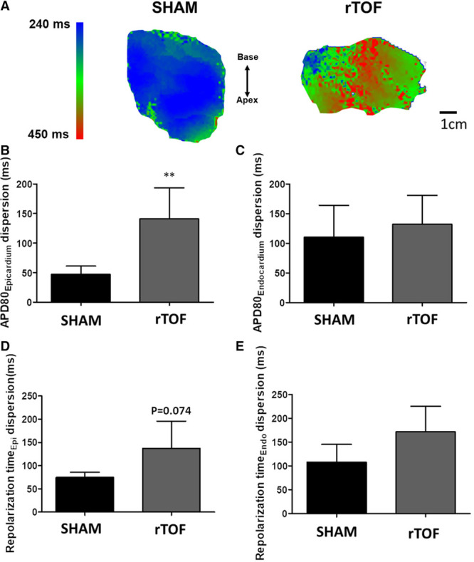
Figure 3.

Dispersion of repolarization in Sham and repaired tetralogy of Fallot (rTOF) left ventricles (LVs). A, Representative epicardial APD80 maps showing heterogeneous action potential duration (APD) distribution in rTOF LVs paced at 1 Hz. B, APD80 dispersion was increased in rTOF compared with Sham in the epicardium but not the endocardium (C). D, There was a trend for an increase in repolarization time dispersion in rTOF LV epicardium but not in the endocardium (E). Data are means±SD. Sham, n=5; rTOF, n=6. **P<0.01.
