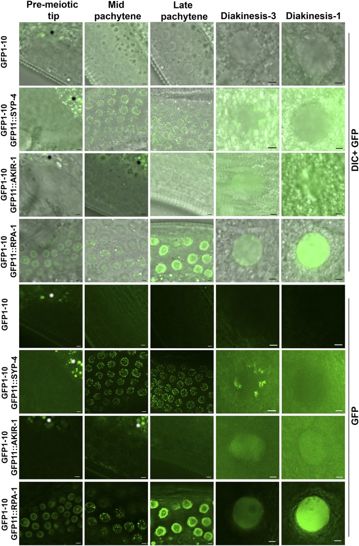
Figure 4.
The split sfGFP approach is suitable for live imaging Expression of GFP11 tagged proteins in the germline expressing GFP1-10 strain leads to localization pattern visible by live imaging. Top is DIC channel and GFP channel, bottom are images taken from the same region just with GFP. Green- live imaging GFP channel. Stages indicated above include the mitotic pre-meiotic tip, and the meiotic stages of mid pachytene, late pachytene, diakinesis (-3 oocyte) and diakinesis (-1 oocyte). Regions marked with * are gut regions that show autofluorescence. All images are slices of 0.8μm close to the mid-section of the nuclei, except SYP-4 which is projection throughout the nuclei. Scale bars 2μm.

