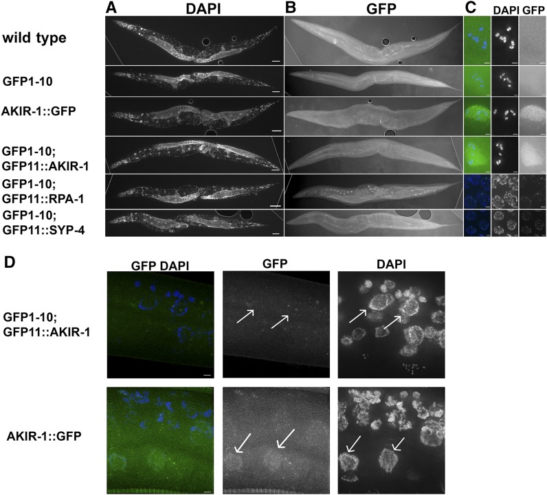
Figure 5.
sfGFP1-10 and GFP11 tagged protein fluorescence is not observed in somatic tissue Images of ethanol fixed worms were taken for both A) DAPI and B) FITC channels. C) Zoom-in images appear to the right of the corresponding worms, and demonstrate the lack of fluorescence in diakinesis (-1 oocyte) nuclei for wild-type and GFP1-10 worms. Representative GFP fluorescence images show diakinesis (-1 oocyte) nuclei for gfp1-10; gfp11::akir-1 and akir-1::gfp strains, mid-pachytene nuclei for gfp1-10; gfp11::rpa-1, and late pachytene nuclei for gfp1-10; gfp-11::syp-4. For all genes tested, fluorescence is not observed in somatic tissue. Dotted lines are where black background was added (externally to that line so a rectangular shape can be made). D) In L3 worm, akir-1::gfp that is expressed from the AKIR-1 promoter and regulated by its 3′UTR localizes to somatic epidermal nuclei, but the same cells in gfp1-10; gfp11::akir-1 do not show GFP fluorescence in these nuclei (representative nuclei are marked by arrows). Scale bars in A and B is 50 µm, in C and D is 2 µm.

