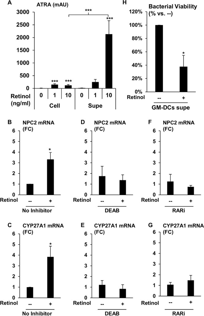
FIG 5.

Activation of monocytes and macrophages with DC-produced ATRA. (A) GM-CSF-derived DCs were treated with the indicated amounts of retinol for 6 h under serum-free conditions, and the amounts of ATRA in the cellular (Cell) and supernatant (Supe) fractions were analyzed via HPLC. Data represent the means ± SEMs of the average area of ATRA peak from HPLC plots (n = 4). Data shown are the average fold change versus control ± SEM (n = 3 to 7). P values by one-way ANOVA. *, P < 0.05; ***, P < 0.001. Expression levels of NPC2 (B) and CYP27A1 (C) were measured by qPCR in primary human monocytes cultured with CM from DCs with or without retinol. From the same experiments, expression levels of NPC2 (D) and CYP27A1 (E) were measured in primary human monocytes cultured with CM from DCs pretreated with DEAB for 20 min prior to addition of retinol. Primary human monocytes were also pretreated with RARi and treated with CM from DC with or without retinol, and mRNA expression levels of NPC2 (F) and CYP27A1 (G) were measured by qPCR. All experimental conditions (panels B to G) were performed in parallel. Data shown are the average fold change versus control ± SEM (n = 3 to 7).
