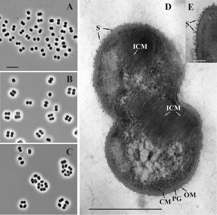
FIG 1.

(A, B, C) Phase-contrast micrographs demonstrating the cell morphology of strain C50C1 in 4-, 7-, and 14-day-old cultures. Bar, 5 μm. (D, E) Electron micrograph of an ultrathin section of a cell. ICM, intracytoplasmic membranes; CM, cytoplasmic membrane; OM, outer membrane; PG, peptidoglycan layer; S, S layer. Bars, 0.5 μm (D) and 0.1 μm (E).
