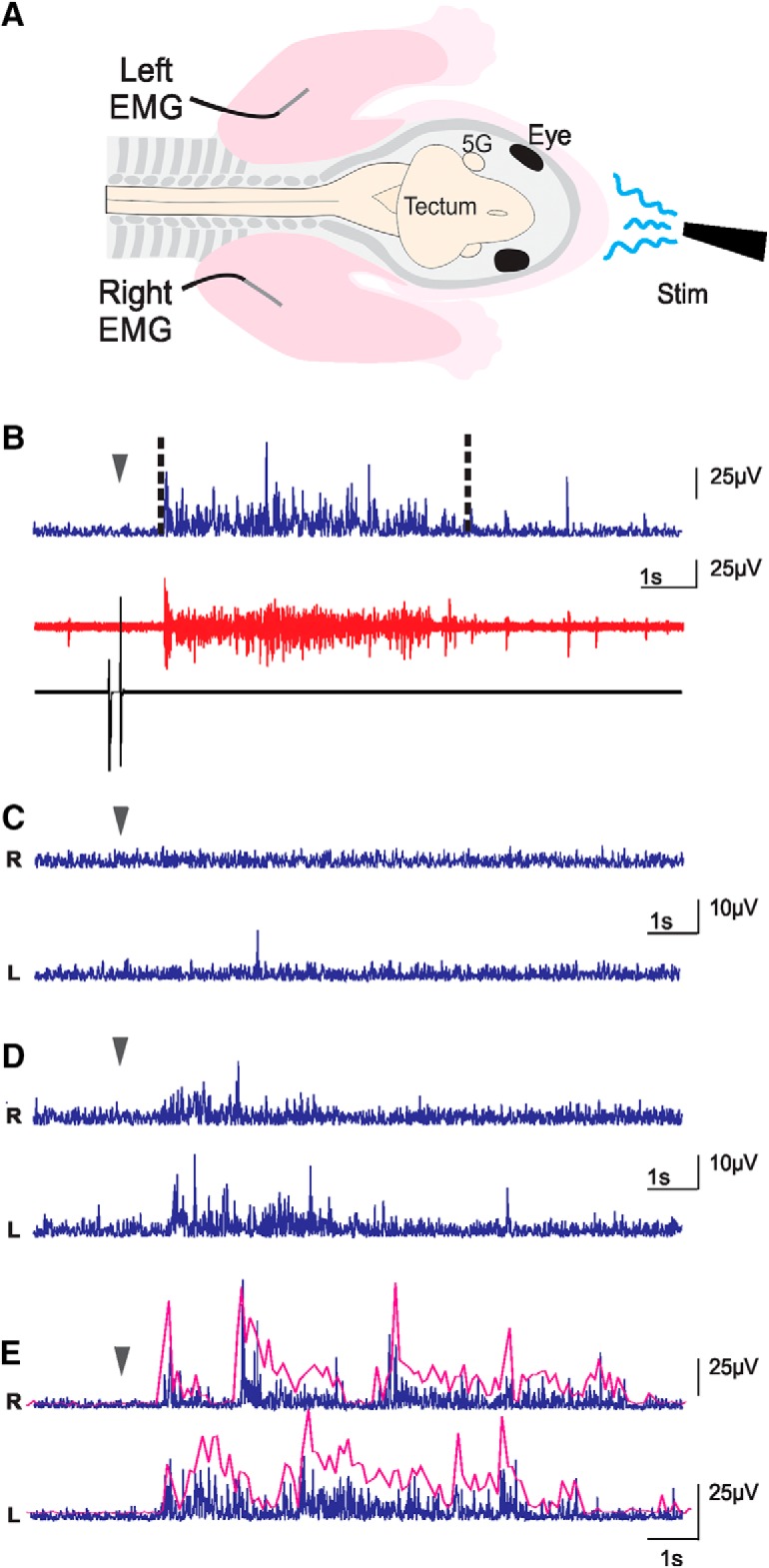
Figure 2.

EMG experiments. A, Schematic representation of the preparations used in EMG recordings. FL were pinned on the bath floor (bath not illustrated) so as to limit movements. Skin was removed on the neck and FL, and EMG electrodes were implanted in triceps muscles. 5G, trigeminal ganglion; Stim, stimulation. B, Muscle activity following a stimulation. Bottom black trace, stimulation artifact produced by the pedal; red trace, raw recording from one EMG; blue trace, same trace as in red, but rectified and with a reduced sampling rate. The dashed lines delimitate the duration of the response used for analysis. C–E, Processed traces exemplifying reactions to stimulation of the left (L) and right (R) triceps muscles of the same animal: no-response (C), uncoordinated response (D), and rhythmic response (E). In B–E, the arrowheads indicate the beginning of the stimulation. The magenta lines in E are envelopes of burst responses highlighting the rhythmical alternation (not to scale with EMG traces).
