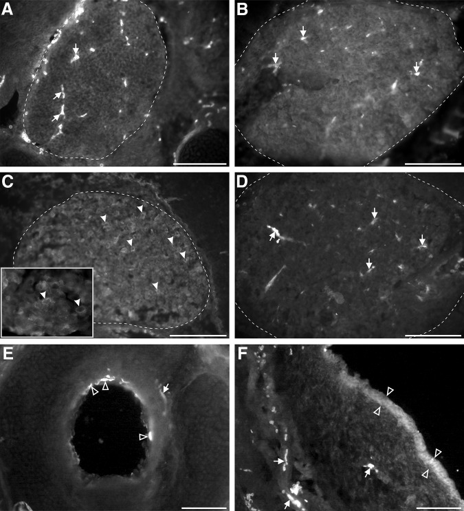
Figure 8.
TRPM8 immunoreactivity in transverse sections of cephalic tissues of newborn opossums. A–D, Trigeminal ganglia (approximately delineated by a dashed line) at P1 (A), P9 (B), and P13 (C, D) processed with (A–C) or without (D) the primary antibody against TRPM8. Labeled cell bodies are present only at P13 (examples pointed by arrowheads in C). The inset in C shows some labeled cell bodies at higher magnification. E, Labeled apical membrane of epithelial cells (empty arrowheads) in the trachea of a P9 opossum. F, Snout from a P9 opossum showing diffuse TRPM8 labeling in the epidermis (between empty arrowheads). Arrows in A, B, D–F point to blood vessels intrinsically fluorescent. Scale bar in F = 100 μm (for A–F).

