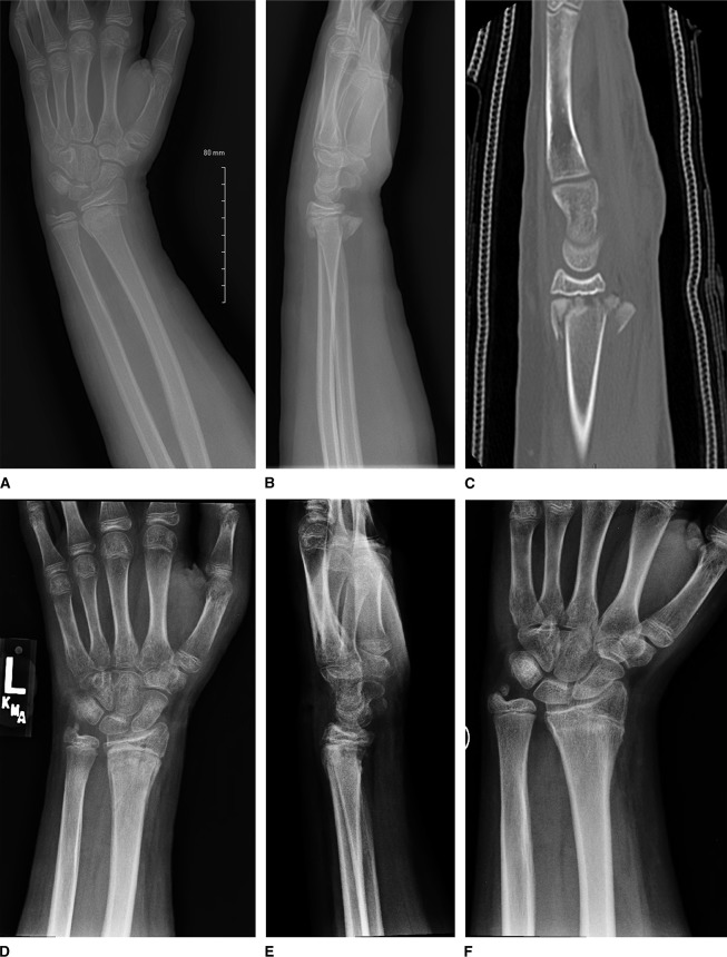
Figure 4.
A, PA wrist radiograph of 12 years, 8 months-year-old patient after fall from standing height while camping. A displaced distal radius physeal fracture is observed in addition to an ulnar styloid fracture. The patient also sustained minimally displaced contralateral distal radius fracture. BMI at the time of injury was 29.7 (98 percentile for age). B, Lateral wrist radiograph of patient (A) demonstrating comminution with loss of radial height and large metaphyseal volar fragment. At the time, patient did not demonstrate any carpal tunnel syndrome. Intraoperatively, the volar fragment was devascularized and required resection to ensure proper reduction and fixation. C, Lateral CT scan of the patient demonstrating comminution and height loss. D, PA wrist radiograph of the patient 4 weeks after closed reduction pin fixation noting full healing of distal radius fracture. E, Lateral wrist radiograph of the patient noting healed fracture and filling in of a previous volar bone defect. F, PA wrist radiograph of the same patient 8 months after fracture fixation, now demonstrating distal radius physeal arrest and ulnar overgrowth. Patient subsequently underwent distal ulna epiphysiodesis.

