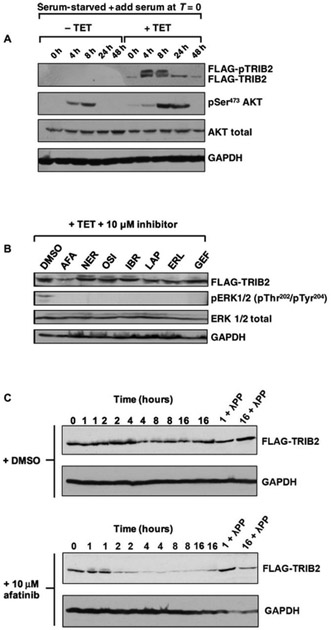
Fig. 3. Afatinib promotes rapid degradation of FLAG-TRIB2 in an inducible HeLa model.
(A) Uninduced (−TET) or TET-induced (+TET) HeLa cells containing a stably integrated FLAG-TRIB2 transgene were serum-starved for 16 hours before the addition of serum and lysed at the indicated times. Whole-cell extracts were blotted with FLAG antibody to detect FLAG-TRIB2 and pSer473 AKT. Total AKT and total GAPDH served as loading controls. (B) A selection of clinically approved kinase inhibitors, including dual EGFR/HER2- and EGFR-specific compounds, were added to TET-induced cells at a final concentration of 10 μM. Stable cells were induced to express FLAG-tagged TRIB2 with TET for 16 hours before inhibitor treatment for 4 hours. Whole-cell extracts were immunoblotted with FLAG, phosphorylated ERK (pERK 1/2), total ERK, or GAPDH antibodies. (C) Stable HeLa cells were incubated with TET for 16 hours and then incubated with DMSO (top panels) or 10 μM afatinib (bottom panels) before lysis at the indicated time. One-hour and 16-hour samples were pretreated with λ protein phosphatase (λPP) before SDS-PAGE. Whole-cell extracts were immunoblotted with FLAG or GAPDH antibodies. Blots are representative of three independent experiments.

