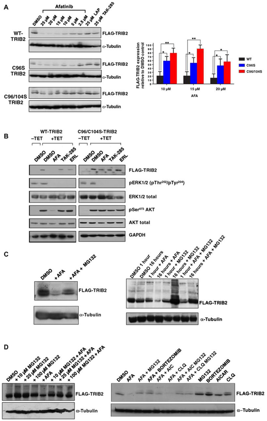
Fig. 4. “On-target” degradation of TRIB2 by afatinib: C96/104S TRIB2 double-point mutant is resistant to degradation.
(A) The indicated concentration of afatinib, lapatinib, or TAK-285 was incubated for 4 hours with isogenic stable HeLa cells expressing FLAG-tagged WT-TRIB2, C96S, or C96/104S TRIB2 (induced by TET exposure for 16 hours). After lysis, whole-cell extracts were immunoblotted with the indicated antibodies. Right: FLAG-TRIB2 abundance was quantified after exposure to 10, 15, and 20 μM afatinib relative to DMSO controls using ImageJ densitometry software. Data are means ± SD from N = 3 independent biological replicates. (B) WT and C96/104S stable HeLa cell lines were subjected to serum block-and-release protocol in the presence (+TET) or absence (−TET) of TET. Subsequently, the indicated compounds (10 μM) were added for 4 hours before cell lysis and immunoblotting with the indicated antibodies. (C) FLAG-tagged TRIB2-expressing HeLa cells were incubated with 0.1% (v/v) DMSO or afatinib (10 μM), in the presence or absence of MG132 (10 μM for 4 hours, left) or at the indicated time points (right) before lysis and processing for immunoblotting. (D) Left: FLAG-TRIB2–expressing stable cells were incubated with the indicated concentration of MG132 in the presence or absence of 10 μM afatinib for 4 hours before cell lysis and immunoblotting. Right: FLAG-TRIB2–expressing stable cells were incubated for 1 hour with MG132 (10 μM), bortezomib (BOR; 10 μM), AICAR (AIC; 1 mM), or chloroquine (CLQ; 50 μM) before the addition of afatinib (10 μM) for an additional 4 hours followed by lysis and immunoblotting with the indicated antibodies. Blots are representative of three independent experiments. *P ≤ 0.05 , **P ≤ 0.01.

