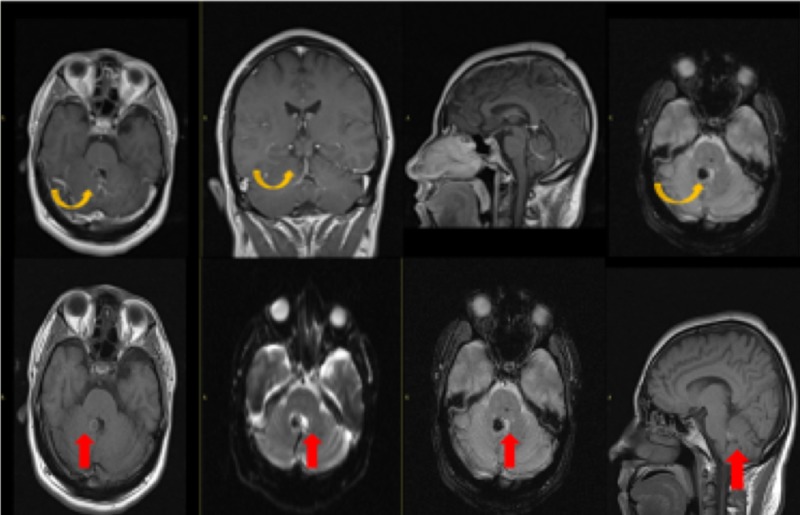
Figure 2. Magnetic Resonance Imaging with Contrast.
Post-contrast images demonstrate a focal area of blooming at the previously seen site of high density on CT at the right medial margin of the fourth ventricle without enhancement on post-contrast (yellow arrow) representing cavernoma. There is also demonstration of caput medusae appearance of small branching veins draining into a single vein adjacent to this lesion (red arrow) suggestive of a developmental venous anomaly (DVA).

