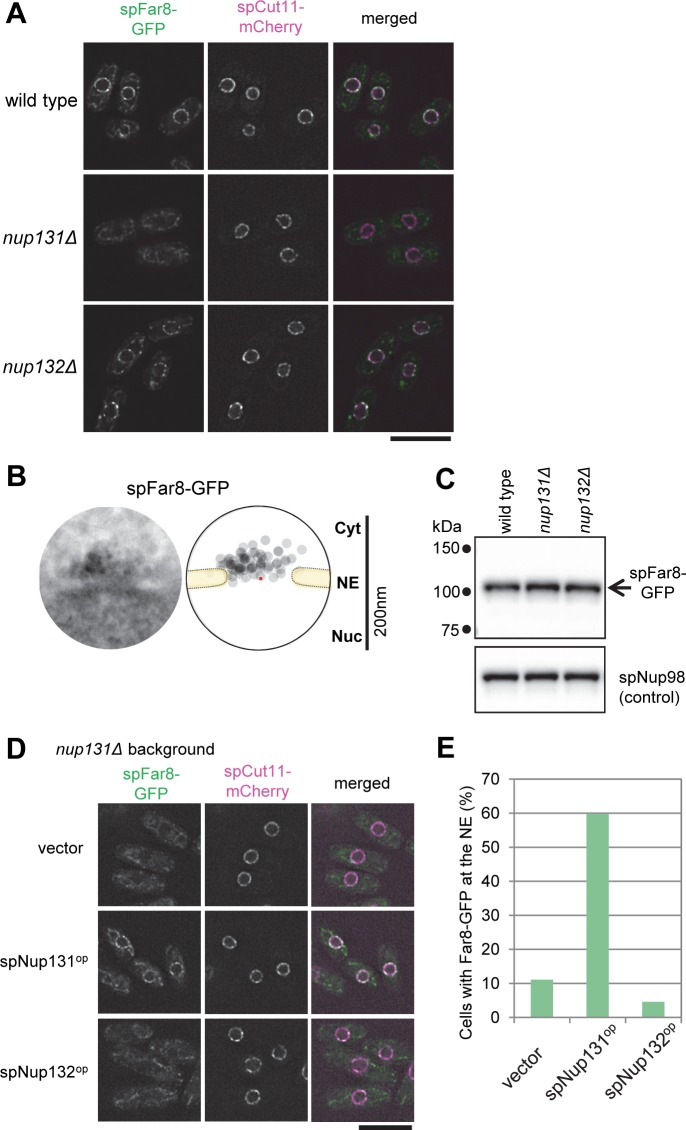
Fig 2. spNup131 is required for spFar8 localization on the cytoplasmic side of the NPC.
(A) Localization of spFar8-GFP in wild type, nup131Δ, and nup132Δ cells. spFar8-GFP was expressed in the indicated strains, and cells exponentially growing in EMM2 liquid medium were observed by FM. Single section images of the same focal plane are shown. spCut11-mCherry was observed as an NPC marker. Scale bar, 10 μm. (B) IEM of spFar8-GFP. For quantitative representation, a montage image of 20 immunoelectron micrographs and a schematic drawing illustrating the distribution of immunogold particles are shown. The red point indicates the pore center. IEM micrographs used for the quantitative analysis are available in S3 Dataset. (C) Western blot analysis of spFar8-GFP. Whole cell extracts were prepared from wild type, nup131Δ, and nup132Δ cells expressing spFar8-GFP and subjected to SDS-PAGE and Western blot analysis. spFar8-GFP was detected with anti-GFP antibody. spNup98 was detected with anti-Nup98 antibody (13C2) as a loading control. The arrow indicates the position of spFar8-GFP. Small dots on the left indicate positions of molecular weight markers. (D) Overexpression of spNup131 and spNup132 in nup131Δ cells expressing spFar8-GFP. spNup131 or spNup132 were overexpressed in nup131Δ cells and localization of spFar8-GFP and spCut11-mCherry was observed by FM. Empty vector was also introduced for a control strain. Single section images of the same focal plane are shown. Scale bar, 10μm. (E) Quantitative analysis of cells exhibiting spFar8-GFP at the NE in the experiment described in (D). Cells observed were: 81, 102, and 130 for vector, spNup131op, and spNup132op, respectively.

