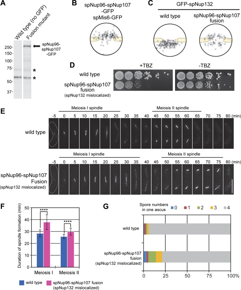
Fig 5. Localization and functional analysis of an spNup96-spNup107 fusion Nup.
(A) Western blot analysis of the spNup96-spNup107-GFP fusion protein. Asterisks represent non-specific cross reactions of the anti-GFP antibody. (B) IEM of the spNup96-spNup107 fusion protein. Immunogold distribution of the projected immunoelectron micrographs is shown. Projection of raw IEM images is shown in S5B Fig. Individual IEM micrographs of 20 NPCs are available in S9 Dataset. (C) IEM of GFP-spNup132 in wild type and spNup96-spNup107 fusion backgrounds. Localization of GFP-spNup132 in a wild type background is taken from the data shown in Fig 1B. Projection of raw IEM images is shown in S5B Fig. Individual IEM micrographs of 20 NPCs are available in S9 Dataset. (D) A cell growth assay in the presence (+TBZ) or absence (-TBZ) of 10 μg/mL TBZ. Five-fold serial dilutions of wild type and spNup96-spNup107 fusion strains were spotted on YES medium containing or lacking TBZ and incubated for 3 days. (E) Time-lapse observation of S. pombe cells (wt or spNup96-spNup107 fusion strains) undergoing meiosis. Cells expressing mCherry-fused α-tubulin (spAtb2) were induced to enter meiosis. The duration of meiosis I and II was determined as the time the spindle was present. Dotted lines show cell shapes. The time 0 indicates the time of the first appearance of meiosis I spindle formation. Representative images are shown as a maximum intensity projection of the serial z-sections (number of cells observed: 32 for wild type and 33 for spNup96-spNup107). (F) Statistical analysis for images obtained in (E). Duration of meiosis I and II. Error bars represent standard deviations. The duration of meiosis I was 28.4 ± 3.0 min in wild type and 37.9 ± 6.3 min in spNup96-spNup107 fusion cells. The duration of meiosis II was 25.8 ± 1.8 min in wild type and 29.8 ± 2.9 min in spNup96-spNup107 fusion cells. Asterisks indicate statistical significance (p < 0.0001) between the indicated strains by Welch’s t-test. Number of cells observed: 32 for wild type and 33 for spNup96-spNup107. (G) Abnormal spore formation was observed in the spNup96-spNup107 fusion background. More than 200 asci were counted for each strain.

