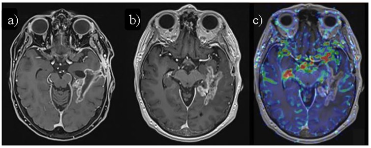
Fig 3. Symptomatic grade 3 radiation necrosis.

a) Axial fat-saturated contrast-enhanced T1-weighted image (CE T1-WI) MRI 4 months after HFRT/TMZ, showing mostly linear enhancement surrounding the surgical cavity in left temporal lobe. b) Axial CE T1-WI of the same patient 2 months later (6 months after HFRT/TMZ), shows increased enhancement with a "soap-bubble" pattern, suggestive of radiation necrosis/pseudoprogression. c) Axial perfusion-weighted cerebral brain volume (CBV) map fused over CE T1-WI shows decreased perfusion values in the enhancing area, further supporting radiation necrosis.
