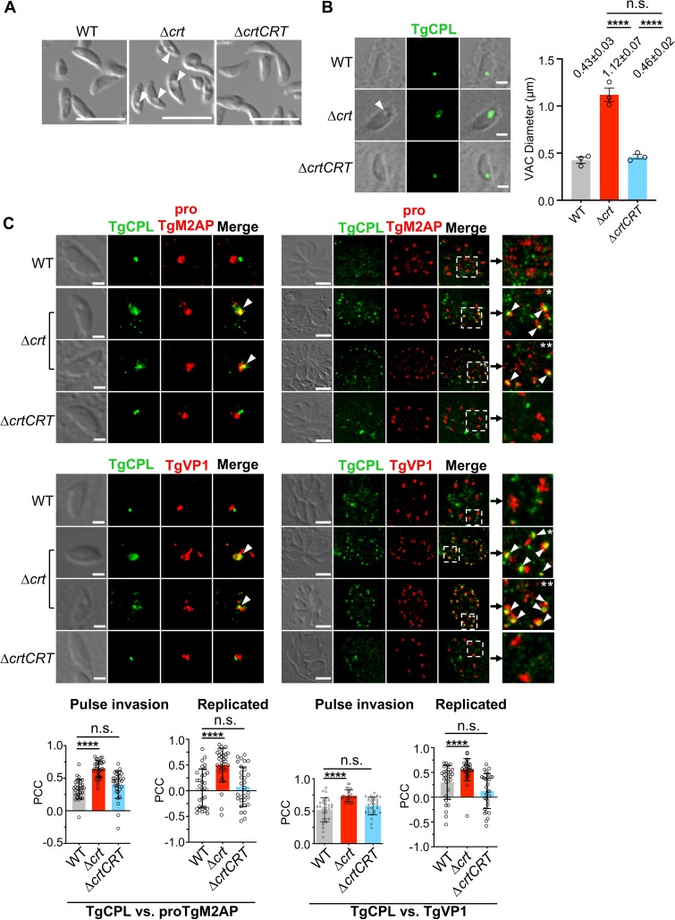
Fig 1. The TgCRT-deficient parasites displayed a swollen vacuole and a disrupted endolysosomal system.
(A) Extracellular Δcrt parasites showed an enlarged concave subcellular structure, indicated by the arrow, under the differential interference contrast (DIC) microscopy. Scale bar = 5 μm. (B) The swollen subcellular structure indicated by the arrow was also observed in pulse-invaded Δcrt parasites and colocalized with a major luminal peptidase of the VAC, cathepsin L-like protease (TgCPL). The TgCPL staining was used to assess the morphology of the VAC. The mean VAC size ± standard error of mean (SEM) was calculated for three independent measurements and is listed in the figure. Statistical significance was determined using unpaired two-tailed Student’s t-test. Scale bar = 2 μm. (C) In pulse-invaded parasites, the TgCPL (VAC marker) and proTgM2AP or TgVP1 (ELC markers) staining were juxtaposed in the WT and ΔcrtCRT strains, but co-localized in the Δcrt strain. During replication, the VAC in WT and ΔcrtCRT parasites became fragmented. However, the aberrant overlap between the VAC and ELC significantly decreased the extent of VAC fragmentation in the Δcrt mutant. TgCPL staining presented as one or more larger and intensely stained puncta, which co-localized with proTgM2AP and TgVP1 (indicated by arrows in the insets). The scale bars in the images of pulse invaded and replicated parasites are 2 μm and 5 μm, respectively. The colocalization of the TgCPL with proTgM2AP or TgVP1 was analyzed by a Coloc2 colocalization analysis software. The Pearson’s correlation coefficient (PCC) of both VAC and ELC staining was measured within 10 individual parasites for each replicate in a total of three replicates and presented as mean ± standard deviation (SD). Statistical significance was determined using unpaired two-tailed Student’s t-test. ****, p<0.0001; n.s., not significant.

