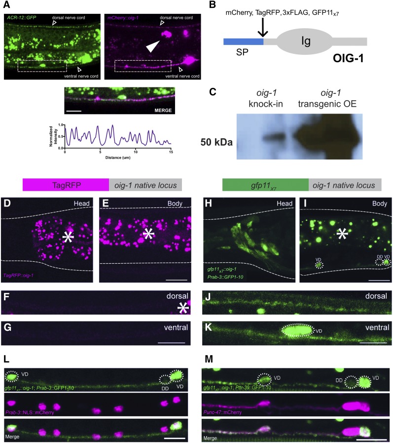
Figure 2.
CRISPR knock-in of GFP11x7 in OIG-1 reveals its intracellular localization in GABAergic motor neurons. (A) Overexpression of mCherry::OIG-1 from a transgenic array (Punc-25::mCherry::oig-1) produces bright puncta (magenta) adjacent to ACR-12::GFP-labeled postsynaptic clusters (green) of AChRs in GABAergic motor neurons (Punc-47::acr-12::gfp). Merge shows ventral nerve cord. Bar, 10 µm. Line scan of mCherry::OIG-1 in the ventral nerve cord (bottom) shows punctate signal. Arrowhead points to coelomocyte. (B) OIG-1 protein showing N-terminal SP, C-terminal Ig (Immunoglobulin) domain, and insertion site for fluorescent and epitope tags (mCherry, TagRFP, 3xFLAG, and GFP11x7). (C) Immunoblot stained for the 3XFLAG tag detects expression of single-copy TagRFP::OIG-1 knock-in and confirms over expression of mCherry::3XFLAG::OIG-1 from multicopy transgenic array. (D–G) TagRFP knock-in at the oig-1 locus (top) does not result in detectable TagRFP::OIG-1 fluorescence. Note absence of TagRFP::OIG-1 signal in head neurons (D), body (E), and dorsal (F) and ventral (G) nerve cords. Asterisks mark autofluorescent granules. (H–K) OIG-1 expression in the nervous system. The gfp11x7::oig-1 knock-in (top) was crossed with the pan-neural transgenic line expressing Prab-3::GFP1-10. A diffuse OIG-1 NATF GFP signal is detected in head neurons (H) and in VD but not DD GABAergic motor neuron cell soma (dashed outlines) in the ventral cord (I). NATF GFP-labeled OIG-1 is detectable in both dorsal and ventral nerve cords (J and K). Asterisk marks autofluorescent granules. (L and M) OIG-1 expression in the nervous system and in GABAergic motor neurons. (L) The gfp11x7::oig-1 knock-in line was crossed with the pan-neural marker Prab-3::gfp1-10 and all neurons labeled with a nuclear-localized pan-neural label, Prab-3::NLS::mCherry. Note OIG-1 NATF GFP signal specifically in VD but not DD cell soma (dashed outlines), nor in additional ventral cord Prab-3::NLS::mCherry-labeled nuclei corresponding to cholinergic motor neurons. (M) The gfp11x7::oig-1 knock-in was crossed with the GABAergic motor neuron-specific marker Pttr-39::gfp1-10 and all GABA neurons were labeled with Punc-47::mCherry. Note the OIG-1 NATF GFP signal in VD but not DD motor neurons (dashed outlines)14. GFP1-10 is cytosolically expressed from the Prab-3::gfp1-10 and Pttr-39::gfp1-10 transgenes, thus indicating that OIG-1 is intracellularly localized at its native expression level. All fluorescent images were obtained from L4-stage larvae. Bar, 10 µm. AChR, acetylcholine receptor; CRISPR, clustered regularly interspaced short palindromic repeats; DD, Dorsal D neuron; GABA, γ-aminobutyric acid; NATF, Native And Tissue-specific Fluorescence; SP, signal peptide; TagRFP, Tag red fluorescent protein; VD, Ventral D neuron.

