Figure 4.
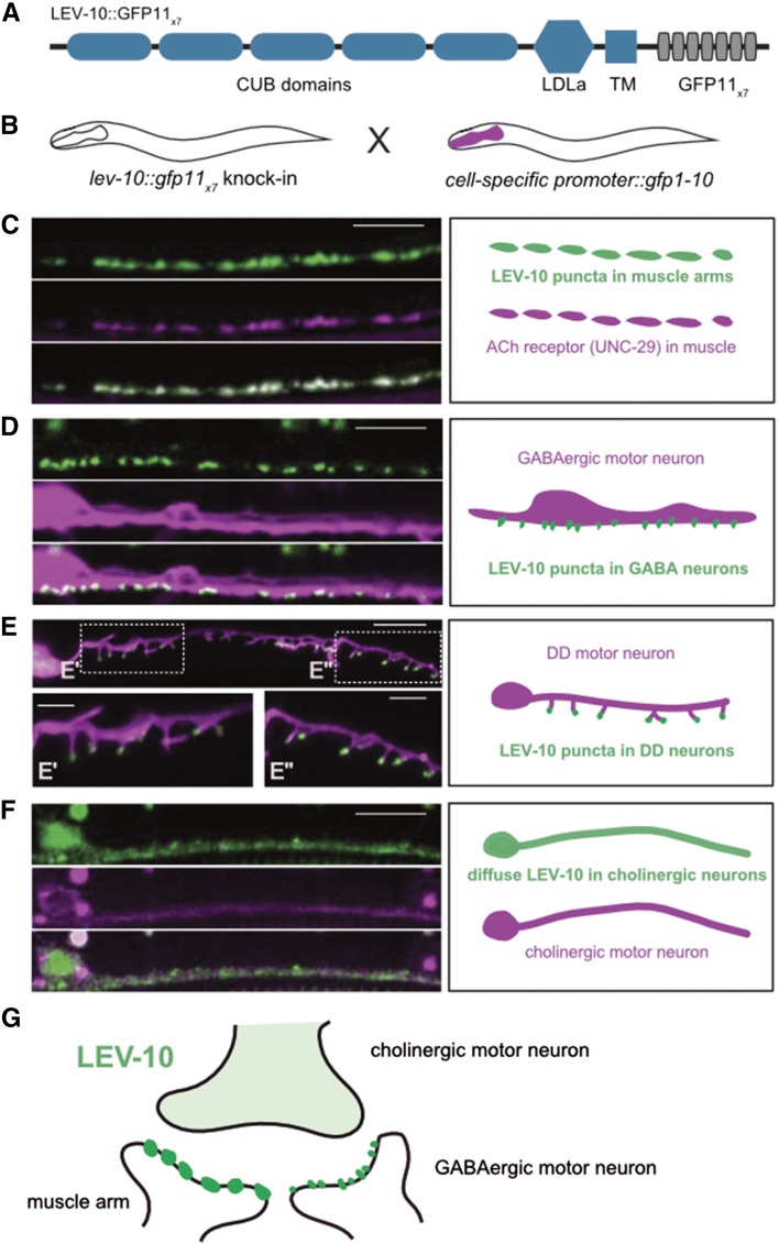
Visualization of LEV-10 NATF GFP signal at cell-specific synapses. (A) Schematic of LEV-10::GFP11x7 showing extracellular complement C1r/C1s, Uegf, Bmp1 (CUB) CUB and low-density lipoprotein receptor domain class A (LDLa) LDLa domains, with TM region and cytoplasmic tail with GFP11x7 insert. (B) Cell-specific labeling strategy. The lev-10::gfp11x7 knock-in strain is crossed with separate transgenic lines expressing GFP1-10 in specific cell types. (C–F) Representative images (left) and schematics (right) of ventral nerve cord region of L4 larvae showing LEV-10 NATF GFP arising from complementation of the lev-10:: gfp11x7 knock-in with cell-specific expression of GFP1-10: (C) Pmyo-3::gfp1-10 (body muscle), (D) Pttr-39::gfp1-10 (DD and VD GABAergic motor neurons), (E) Pflp-13::gfp1-10 (DD GABAergic motor neurons) with superresolution images with insets (E’ and E’’) showing localization of LEV-10 NATF GFP to tips of postsynaptic DD dendritic spines, and (F) Pacr-2::gfp1-10 (cholinergic motor neurons). Pmyo-3::unc-29:TagRFP marks ACh receptors in body muscle in (C), Punc-47::mCherry labels GABA neurons in (D), Pflp-13::LifeAct::mCherry marks DD neurons in (E), and Punc-4::mCherry labels cholinergic motor neurons in (F). Bars, 5 (C–F) and 2 µm (E’ and E’’). (G) Schematic showing distribution of LEV-10 NATF GFP at a dyadic synapse of a presynaptic cholinergic motor neuron with postsynaptic body muscle and GABAergic motor neuron in the ventral nerve cord. ACh, acetylcholine; DD, Dorsal D neuron; GABA, γ-aminobutyric acid; NATF, Native And Tissue-specific Fluorescence; TagRFP, Tag red fluorescent protein; TM, transmembrane; VD, Ventral D neuron.

