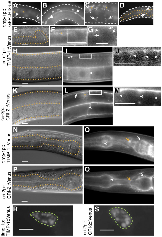
Figure 3.
Expression and localization patterns of TIMP-1 and CRI-2. (A–D) Expression of the transcriptional reporter GFP::HIS-58 under the control of the putative endogenous timp-1 5′ cis-regulatory region. The bkcSi4[timp-1p::GFP::his-58] allele was used. In young adults, signals were detected in epidermal cells (arrows, A), seam cells (arrows, B), the lateral region of the vulva (arrows, C), and a subpopulation of neuronal cells in the nerve ring (arrows, D). DIC (E, H, K, N, and P) and fluorescence (F, G, I, J, L, M, O, and Q) images of L3-stage larvae (E–G) and young adults (H–Q). Images of the posterior gonads (E–M) and pharynges (N–Q) of worms with timp-1(tk71);bkcSi1[timp-1p::timp-1::Venus] (E–J, N, and O) and bkcSi7[cri-2p::cri-2::Venus] (K–M, P, and G). (G, J, and M) display, at a higher magnification, the squares enclosed by the white boundaries in (F, I, and L). The contrast was enhanced to visualize the basement and plasma membrane localization of the signals. (E–Q) White arrows, white arrowheads, and orange arrows indicate the basement membrane, plasma membrane, and nerve ring, respectively. Fluorescence images of the coelomocyte of (R and S) young adult worms with bkcEx1[timp-1p::timp-1::Venus] (R) and bkcEx2[cri-2p::cri-2::Venus] (S). The white, orange, and green dotted lines outline the worm, the gonad or pharynx, or coelomocyte, respectively. The orange asterisks indicate autofluorescence signals (A, C, and F) or signal cross talk from the coelomocyte (L), a scavenger cell that takes up secreted molecules such as CRI-2::Venus. In all panels, the anterior region of the gonad is to the left and its dorsal region is at the top of the image. Three independent timp-1p::GFP::his-58 transgenic lines exhibited similar expression patterns. Bars, 10 μm.

