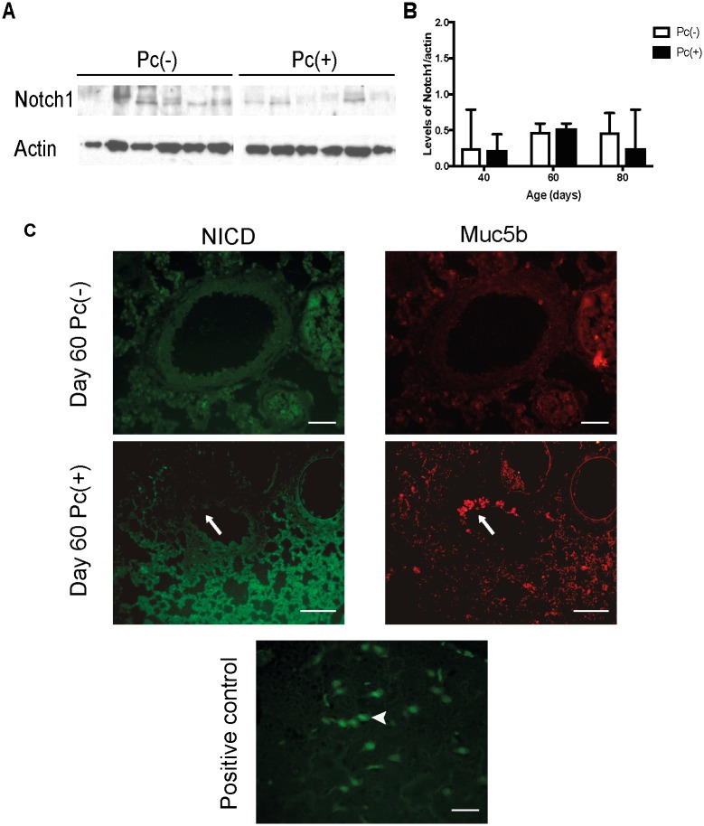
Fig 6. Components of Notch pathway in the whole lung and distal airway.
Protein levels of Notch1 were determined in the whole lungs of Pc(-) and Pc(+) groups, at different days of infection by western blot. Localization of activated Notch intracellular domain (NICD) was determined in distal airway by IIF with antibodies anti-activated Notch1 (NICD) and anti-Muc5b. (A) Representative experiment of protein levels of Notch1 in the day 40 of infection. (B) Densitometric analysis of the Notch1 western blots. Data are shown as median and interquartile range. (C) Representatives images of NICD and Muc5b of Pc(-) and Pc(+) groups at day 60 of infection, and positive control (brain). Scale bars = 25 μm and 100 μm, magnification 40x and 10x, respectively. Arrows indicate immunoreactive cells to Muc5b but not to NICD. Arrowhead in positive control (brain tissue) shows an immunoreactive cell to NICD. For all experiments, n = 6.

