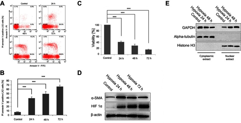
Figure 1.
Hypoxia exposure affects the activation of LX2 cells and increases cell apoptosis and GAPDH nuclear translocation. (A and B) Cell apoptosis was detected using annexin V-FITC/PI double staining and flow cytometry (A), and bar graphs (B) show the effect of hypoxia on apoptosis. (C) Cell viability was measured using the MTT assay after hypoxia. (D and E) Western blots showing the effects of hypoxia on the activation of LX2 cells (D) and the accumulation of nuclear GAPDH (E). Untreated samples served as controls.
Note: Data are presented as the mean ± SEM. n.s., not significant, *p<0.05, **p<0.01, and ***p<0.001, n=3.
Abbreviations: GAPDH, glyceraldehyde-3-phosphate dehydrogenase; α-SMA, α-smooth muscle actin; HIF-1α, hypoxia-inducible factor 1α.

