Figure 1.

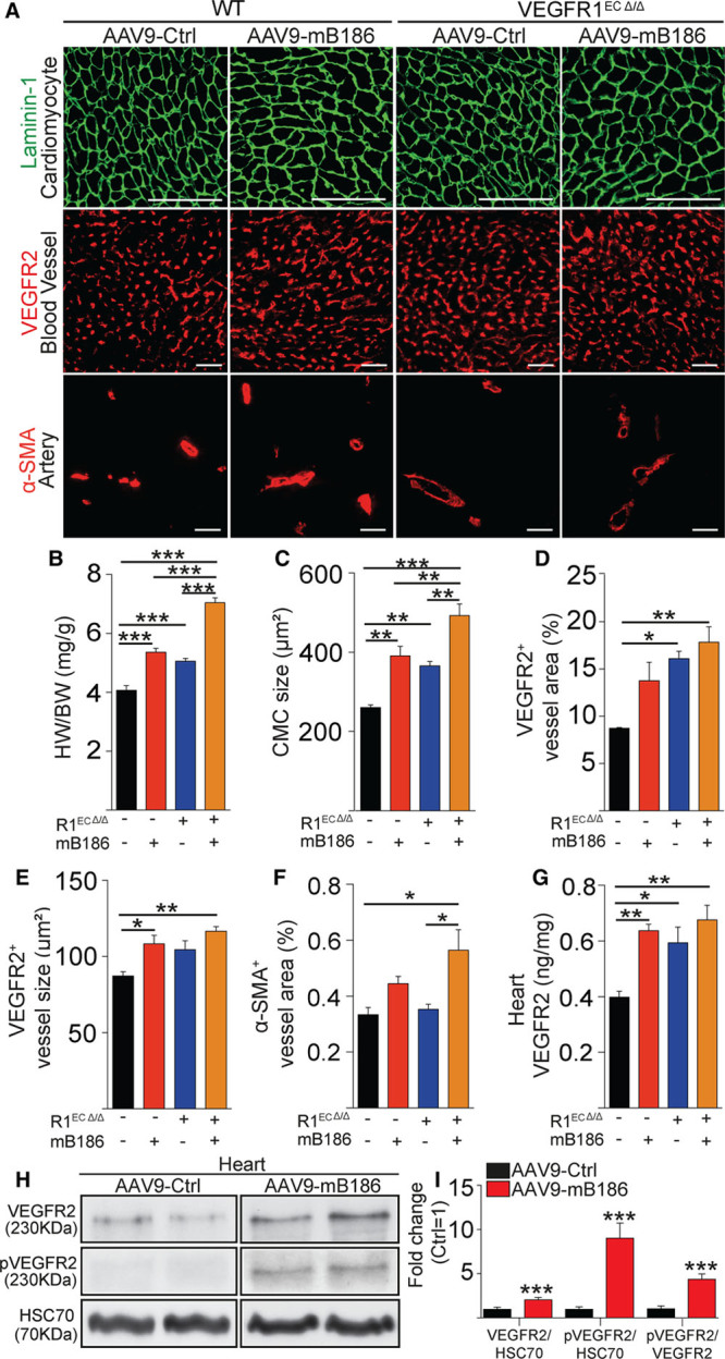
VEGFR1 deletion from endothelial cells induces angiogenesis and cardiomyocyte hypertrophy. A, Representative images of staining for cardiomyocytes (Laminin-1), blood vessels (VEGFR2), and arteries (α-SMA) in AAV-VEGFB186–treated or VEGFR1-deleted hearts (R1ECΔ/Δ). B and C, Quantification of heart weight normalized to body weight and cardiomyocyte size (in µm2). D through F, Quantification of blood vessel area, average capillary size (in µm2), and α-SMA area. G, VEGFR2 protein concentration (in ng/mg) in the heart. H, Western blots of total VEGFR2 and phosphorylated VEGFR2 in VEGF-B–overexpressing and wild-type hearts. Heat shock cognate 70 (HSC70) was used as a loading control. I, Quantification of the Western blot signals is shown as fold change compared with AAV-Ctrl treatment. Data are mean±SEM. AAV9 indicates adeno-associated viral vector serotype 9; CMC, cardiomyocyte; Ctrl, control; HW/BW, heart weight/body weight; mB186, mouse VEGF B-186; pVEGFR2, phosphorylated VEGFR2; α-SMA, α-smooth muscle actin; VEGF-B, vascular endothelial growth factor B; VEGFR2, vascular endothelial growth factor receptor 2; and WT, wild type. Two-way ANOVA (Holm-Sidak test) and Student t test were used, as appropriate; *P<0.05, **P<0.01, ***P<0.001 (N=4 per group). Scale bar, 100 µm.
