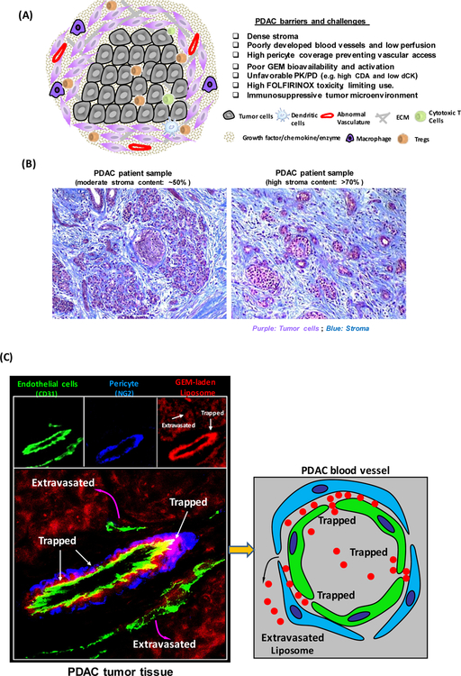
Figure 1.
(A) Schematic to show the barriers and challenges that are responsible for failed chemotherapy in PDAC, including as a result of an abundant dysplastic stroma, which serves as a physical and biological barrier, including the immunosuppressive tumor microenvironment. (B) Trichrome staining of PDAC tissue sections. Moderate (~50%) and high (>70%) stroma content PDAC tumors were shown. Blue: stroma, e.g. collagen. Purple: Tumor cells. (C) Figure 1C demonstrates that PDAC tumor develops a dense stromal barrier, which blocks the vascular access of IV-injected red fluorescent liposomes. Higher level magnification was provided to show the localization of the liposomes in the tumor in relation to endothelial cells (CD31) and pericyte (NG2) fluorescent markers. The fluorescence microscopy image obtained from PDAC tumor site showed a region of a blood vessel where pericytes were trapping some liposomes just beyond their point of egress from the vascular fenestrations [23].

