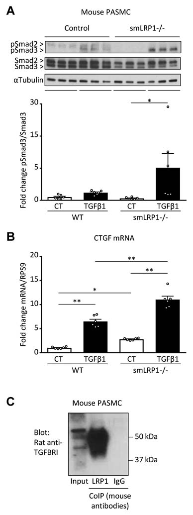
Figure 2. LRP1 regulates TGFβ1 pathways in PASMC.
(A-C) PASMC were isolated from the main pulmonary artery of WT (n=2) or smLRP1−/− mice (n=2). (A) Phosphorylated and total protein expression of Smad2 (60 kDa) and Smad3 (52 kDa) expression was evaluated by western blot (n=6 experiments). (B) CTGF mRNA expression was evaluated by PCR (n=6 experiments). (C) Total cell lysates from WT mouse PASCM were subjected to coIP with anti-LRP1 or IgG (both mouse antibodies) and immunoblotted for TGFBRI (rat antibody) (n=4 experiments). All values are expressed as mean ± SEM; *p<0.05; **p<0.01.

