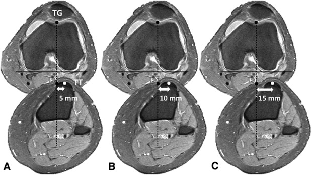
Fig. 1 A-C.

The MRI measurement technique for TT-TG distance is demonstrated. The black dot represents orientation of the trochlear groove (TG) designated by the deepest cartilaginous point on the image. The tibial tubercle (TT) is denoted by the white dot as determined from the central insertion site of the patellar tendon. The horizontal continuous line illustrates the posterior intercondylar line. The vertical dashed line renders the intersection of the intercondylar line per position of the TG, which is used to calculate distance in reference to the TT. (A-B) This is a typical range for what constitutes a normal TT-TG distance [58, 75, 83]; (C) this resembles an abnormal TT-TG distance that may receive limited benefit from rehabilitation in the presence of PFI [83].
