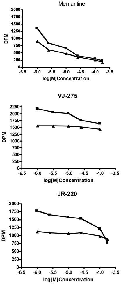
Fig. (1).
Typical data from the “differential” molecular screen showing inhibition of [3H]MK801 binding in the presence (upper curves) and absence (lower curves) of potentiation by 100μM spermidine. The data are presented as untransformed DPM rather than as % specific binding, because the upper curves represent inhibition of time-dependent potentiation of [3H]MK801 binding by spermidine.

