Figure 3.
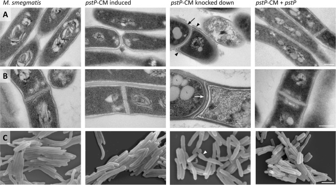
Knockdown of the pstP operon led to changes in the cell wall and cell shape. Knockdown for 30 hours led to changes in pstP-CM compared to wild type M. smegmatis, and some of these changes were reversed in the complemented strain pstP-CM + pstP. Cells were imaged by transmission electron microscopy (A,B) and scanning electron microscopy (C). PstP knockdown led to thicker cell wall (black arrow), thicker septum (white arrow), and bulging cells with lysis (white arrowhead). These defects were complemented by reintroduction of pstP. Knockdown of the pstP operon also led to rough cell surface (black arrowhead) and lipid bodies (L) that were partially complemented by reintroduction of pstP, and reduced cell length (quantified in Fig. S8) that was not complemented by reintroduction of pstP. Images within a single row are shown at the same scale, which is indicated by a bar in the right hand image: 200 nm for TEM (A,B) and 2.5 μm for SEM (C). Images are representative of at least twelve fields of view: additional fields for transmission electron microscopy are presented in Figs S6 and S7, and cell lengths for 100 cells are presented in Fig. S8.

