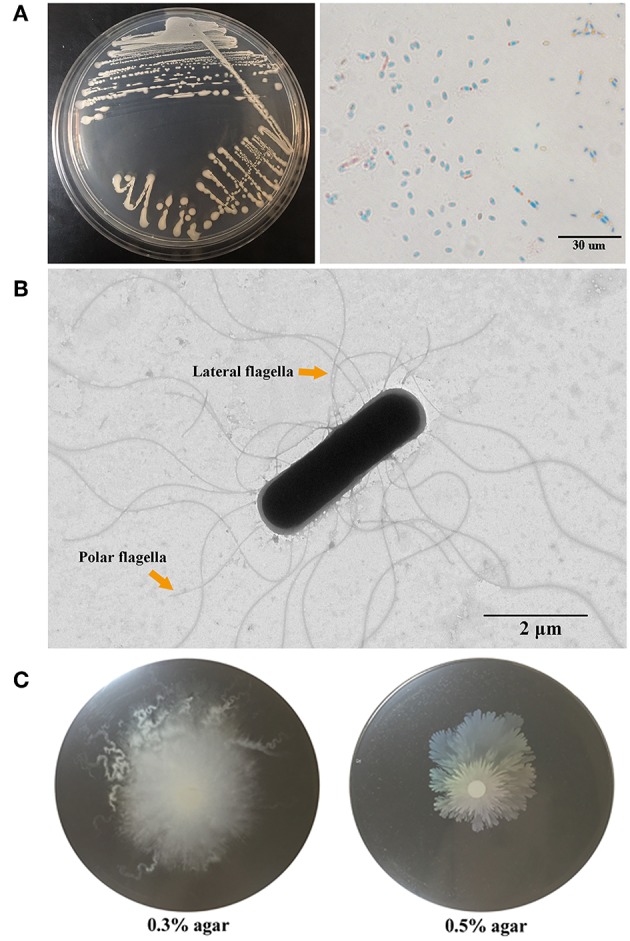
Figure 1.

Morphology and motility of G7. (A) G7 colonies on Marine 2216E plate (left) and micrograph of malachite green and safranine stained spores of G7 (right). (B) G7 was observed with a transmission electron microscope, the arrows indicate polar and lateral flagella. (C) G7 suspension was spotted onto the center of marine 2216E plates containing 0.3 or 0.5% (w/v) agar, and the plates were incubated at 28°C for 24 h.
