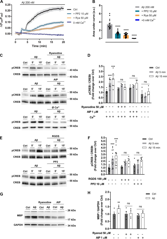
Fig. 4. Aβ-induced intracellular Ca2+ increase and CaMKII activation upregulate MBP expression in oligodendrocytes.
a Cells were exposed to Aβ oligomers and Ca2+ levels were measured in Ca2+-containing extracellular buffer in the presence of PP2 (SFK inhibitor) or the RyR inhibitor ryanodine, and in Ca2+-free extracellular buffer (n = 4). b Bars represent the mean ± SEM of Ca2+ increases, quantified as the area under the curve in treated oligodendrocytes. c, d Western blot of CREB phosphorylation (pCREB) in cells treated with Aβ 200 nM for 5 or 15 min, and preincubated with ryanodine or AIP (CaMKII inhibitor). pCREB levels were also analyzed in the presence or absence of Ca2+ (n = 3; lower panel). e, f Cells were preincubated with RGDS (integrin inhibitor) or PP2 (SFK inhibitor) and treated with Aβ 200 nM for 5 or 15 min. CREB activation was detected and analyzed by western blot (n = 6). g, h Cells with ryanodine or AIP, were treated with Aβ 200 nM for 24 h and MBP expression levels were examined by western blot. Data are represented as means ± S.E.M and were analyzed by one-way ANOVA followed by Holm-Sidak’s multiple comparisons test (a, b) and two-way ANOVA followed by Bonferroni posttest (d, f, h). *p < 0.05, **p < 0.01, ***p < 0.001 compared to non-treated cells

