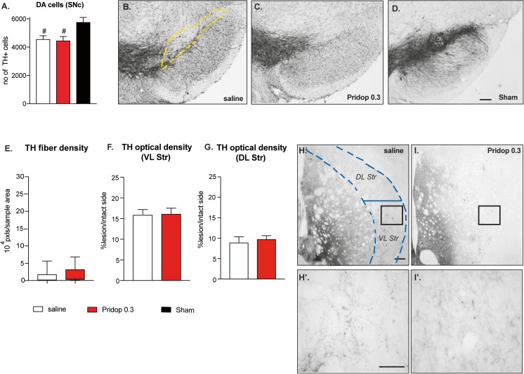
Fig. 5.
Pridopidine is devoid of neurorestorative effects in 6-OHDA-lesioned mice that lack S1Rs. (A) Stereological counts of TH-positive cells in the SNc in S1R KO mice and (B-D) low-magnification photomicrographs of nigral sections immunostained for TH (the dashed yellow line outlines the area included in the analysis). Scale bar, 100 μm. Data in A show the total number of cells from the SNc ipsilateral to the lesion (counted on 7 sections per animal throughout the SNc). One-way ANOVA p < 0.0001, F(2,26) = 41.73; post hoc Bonferroni test; p < 0.05 number sign versus sham group. (E) Image segmentation analysis of TH-positive fibers as performed at high magnification on random sample areas through the lateral striatum (encased by dashed blue lines). Values are expressed as fiber pixels/sample area, whiskers encompass the entire range of values in each group, the median is shown as a horizontal line. Mann–Whitney test, p = 0.084. (F-G) Optical density (O.D.) of TH immunostaining over the entire cross-sectional area of ventrolateral (F) and dorsolateral (G) striatum. O.D. values from the side ipsilateral to the lesion are expressed as a percentage of those from the contralateral intact side in each animal. (F) Ventrolateral (VL) striatum, unpaired t test, p = 0.516; (G) Dorsolateral (DL) striatum, p = 0.219. (H-I′) Representative photomicrographs of TH-immunostained striatal sections from a saline-treated (H, H′) and a pridopidine-0.3 mg/kg treated S1R KO mouse (I, I′). Scale bar, 200 μm

