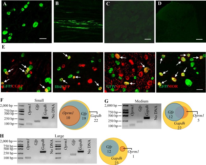
Fig. 1.
GFP-labeled HSV is limited in the ipsilateral L5 DRG neurons and their fibers on day 7 after microinjection of HSV-GFP into the unilateral L5 DRG. (A–D) GFP expression in the L5 DRG (A) and L5 spinal nerve (B) on the ipsilateral side. No GFP expression in the contralateral L5 DRG (C) and ipsilateral L5 spinal cord dorsal horn (D). Scale bars, 50 μm. (E) GFP-labeled neurons are positive for CGRP (43.5%), IB4 (31.7%), NF200 (26.8%), and MOR (38.3%) (arrows). n = 3 biological repeats (3 rats). Scale bars, 50 μm. (F–H) Co-expression of Oprm1 mRNA with Gfp mRNA in small (F) and medium (G) DRG neurons, but not in large DRG neurons (H). A representative gel of a single-cell real-time RT-PCR assay (left) and a Venn diagram of Oprm1 mRNA-, Gfp mRNA-, or Gapdh mRNA-expressed neurons in the L5 DRG (right) are shown in (F), (G), and (H)

