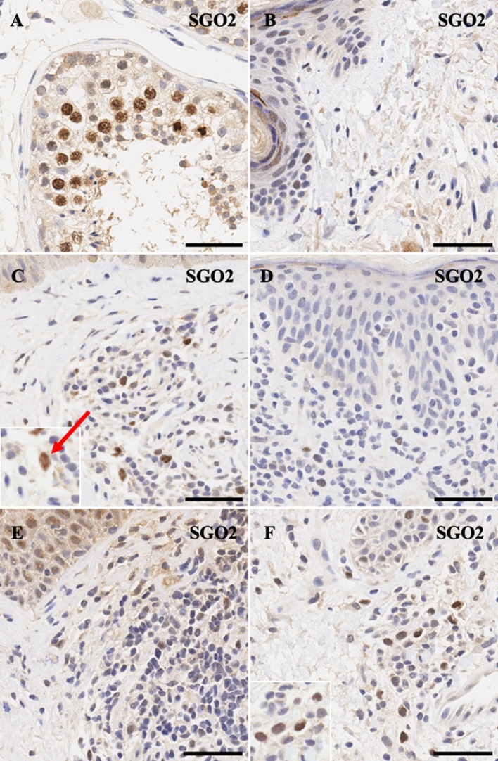
Figure 4.
Immunohistochemistry staining of SGO2 in (A) normal human testis (positive control), (B) normal skin (C) stage IIA MF lesional skin, (D) CD8+ MF lesional skin, (E) Sézary Syndrome (F) peripheral T-Cell Lymphoma. Scale bars are 50 μm. Nuclear staining in malignant lymphocytes is highlighted (red arrow).

