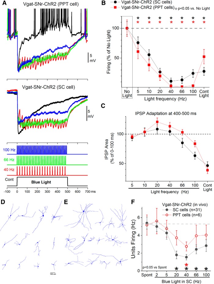
Figure 5.
Postsynaptic effects of activating GABAergic fibers with optogenetics. A, Example intracellular whole-cell responses obtained in two different neurons after activating GABAergic fibers that express ChR2 with continuous pulses or trains of 1 ms pulses of blue light. The GABAergic fibers originate in SNr and the recorded cells are in PPT (top) or superior colliculus (bottom). Each trace is an average of six trials. B, Effect of blue light on the firing of neurons recorded intracellularly for the different pathways: SNr-to-PPT and SNr-to- superior colliculus (SC). Firing was spontaneous (resting Vm) or induced with positive current injection. C, Adaptation of the hyperpolarizing responses (IPSPs) caused by activating GABAergic fibers. The area of hyperpolarization measured at the end (400–500 ms) of each type of optogenetic stimulus is plotted as a percentage of the area at the onset (0–100 ms) of the stimulus to reveal the amount of adaptation of the response as a function of the stimulus type. D, Cells (n = 12) recorded in PPT that were inhibited by SNr GABAergic afferents. E, Cells (n = 13) recorded in superior colliculus that were inhibited by SNr GABAergic afferents. F, Units recorded in the superior colliculus and PPT of urethane-anesthetized Vgat-SNr-Chr2 mice during application of blue light trains in the superior colliculus. Blue light trains delivered in superior colliculus at 40 Hz inhibit cells in PPT, presumably through antidromic activation of SNr cells that project to both superior colliculus and PPT.

