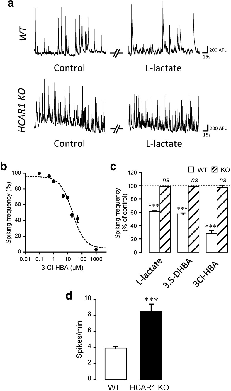
Figure 3.
Effect of neuronal activation in neuronal calcium spiking activity. a, Representative traces of calcium spiking in control or 5 mm l-lactate-containing solutions, for both WT and HCAR1 KO neurons. b, The effect of the HCAR1 agonist 3Cl-HBA on spontaneous calcium activity of neurons was concentration dependent, with an IC50 value of 21.5 ± 6.1 μm (n = 177 neurons from 23 experiments). The IC50 value was obtained by nonlinear curve fitting using the Levenberg-Marquardt algorithm. c, Effect of HCAR1 activation on calcium spiking frequency from WT and HCAR1 KO neurons with 5 mm l-lactate (WT: n = 66 cells, 16 experiments; HCAR1 KO: n = 81 cells, 16 experiments), 1 mm 3,5-DHBA (WT: n = 57, 16 experiments; HCAR1 KO: n = 74 cells, 12 experiments), or 40 μm 3Cl-HBA (WT: n = 31 cells, 3 experiments; HCAR1 KO: n = 32 cells, 5 experiments). Spiking frequency is shown as a percentage of activity measured in the control condition. The effect of HCAR1 activation was reversible in all experiments (data not shown). Significance is shown compared with control and among conditions. d, Comparison of basal spontaneous spiking frequencies of neurons from WT (n = 16) and HCAR1 KO (n = 16) mice. ***p < 0.001, ns, not significant.

