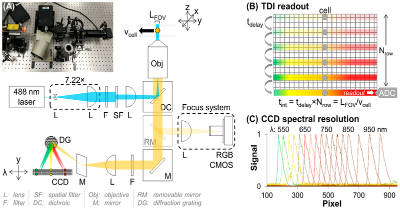
Figure 1.
(A) Picture and optical diagram of the TDI-SFC. A beam expander and epi-illumination lens produced widefield excitation in the flow cell from the 488 nm laser. Fluorescence emission was filtered and passed to a spectrograph that spectrally dispersed and focused the emission onto a back-illuminated CCD. If engaged, a removable mirror provided imaging for system focusing using a CMOS camera. (B) Illustration of TDI mode. The CCD’s delay time, which sets the shift rate, is matched with the cell’s velocity through the flow cell, providing integrated readout of the cell’s fluorescence. (C) Cell’s fluorescence spectrally resolved via the spectrograph.

