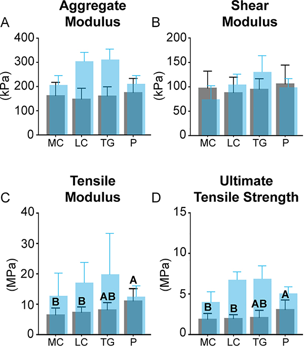
Fig. 4.
Mechanical characterization: mechanical properties of the fetal ovine medial condyle (MC), lateral condyle (LC), trochlear groove (TG), and patella (P) are shown in gray. Historical values of biochemical content of juvenile ovine cartilage from the same regions are shown in translucent blue [19]. No regional differences in aggregate modulus (A) or shear modulus (B) were observed. The P was stiffest (C) and strongest (D) in tension. Topographical mechanical data are available in Table 2. (For interpretation of the references to colour in this figure legend, the reader is referred to the web version of this article.)

