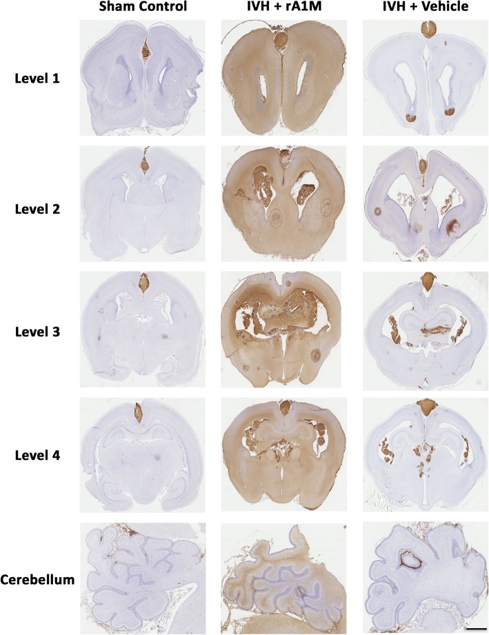
Fig. 2.
Cerebral and cerebellar A1M distribution following i.c.v. administration of rA1M in rabbit brain following IVH. IHC labeling of A1M was performed to investigate the distribution of i.c.v. administrated rA1M. Rabbit pups with confirmed IVH received i.c.v. injections of either rA1M (IVH + rA1M) or Vehicle (IVH + Vehicle) and were euthanized at 72 h of age followed by saline and freshly prepared 4% PFA perfusion. Brains were prepared and sections from control animals (Sham Control, no bleeding as confirmed with high-frequency ultrasound), IVH + rA1M and IVH + Vehicle animals, at the levels of rostral forebrain (Level 1), caudal forebrain (Level 2), rostral midbrain (Level 3), caudal midbrain (Level 4) and cerebellum were immunolabeled against A1M as described in the “Materials and methods” section. Microscope analyses were performed on a wide-field Olympus microscope (IX73) and slide scanning were performed on a Hamamatsu NanoZoomer 2.0-HT Digital slide scanner: C10730. Scanning was performed with a 40x magnification lens. Scale bar indicates 5 mm and is applicable for all images

