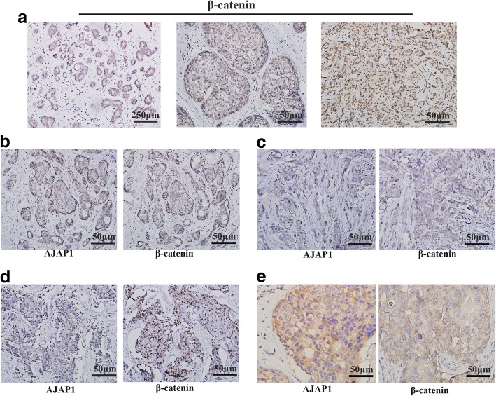
Fig. 2.
Immunohistochemical expression of β-catenin and AJAP1 in breast tissue slides. a β-catenin expression in normal breast cancer tissues, ductal carcinoma in situ and invasive ductal carcinoma (magnification, × 40, × 200 and × 200). b-e Different expression patterns of AJAP1 and β-catenin in breast cancer tissues. Micrographs showing low expression (c, d) and high expression (b, e) AJAP1 cytoplasm (b, c, d) and membrane (e) expression and negative (c) and positive (b, d, e) β-catenin membrane(e), cytoplasm (b, c) and nuclear expression(d) through the immunohistochemical staining of breast cancer specimens (magnification, × 200)

