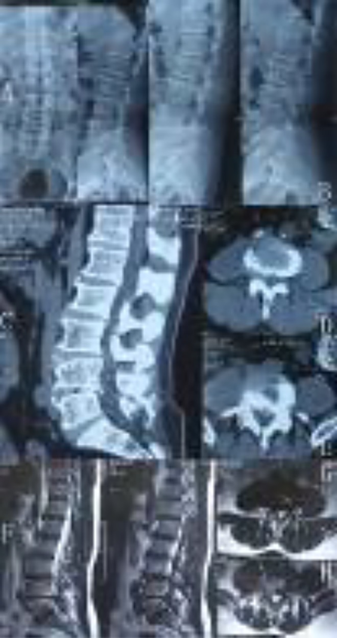
Figure 1.

Preoperative film material. (A) AP (anteriorposterior) and lateral X-ray film. (B) Overextension and overflexion X-ray film. (C) CT (computed tomography) scan film. (D): L34 layer scan film shows that there is large disc herniation in left part of the canal. (E) L45 layer scan. There is a large and marginal calcified disc herniation. (F) Lumbar MRI (magnetic resonance imaging) scan film. (G) There is a large disc herniation at the L34 layer. (H) It is a large and marginal calcified disc herniation at the L45 layer.
