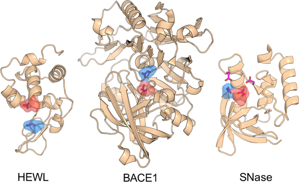
Figure 1.

Crystal structures of HEWL (PDB ID 2LZT), BACE1 (PDB ID 1SGZ) and SNase (PDB ID 3BDC). The structures of BACE2 and CatD are very similar to BACE1 and not shown here. The active-site residues are represented by the stick model. The catalytic dyads in HEWL and BACE1 as well as the two hydrogen bonded catalytic residues in SNase are highlighted by surface rendering. The proton donor and nucleophile are colored blue and red, respectively.
