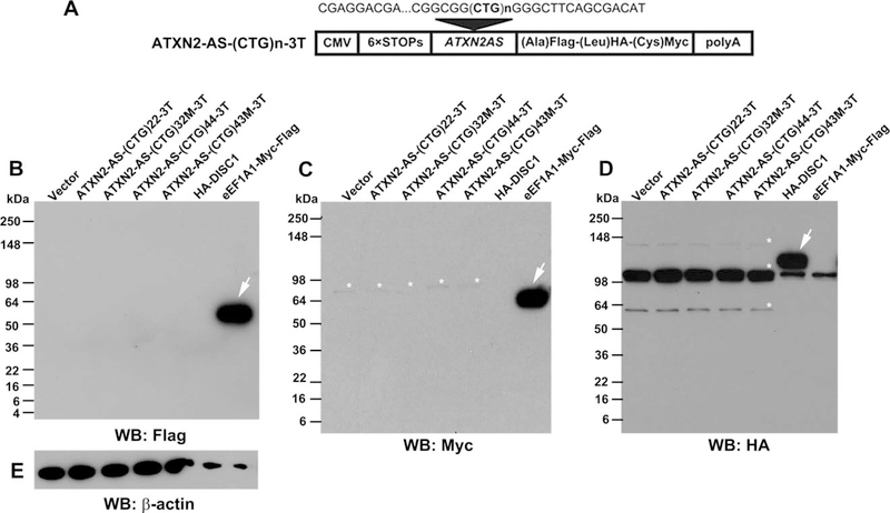
FIGURE 6:
Non-ATG initiated (RAN) translation does not contribute to the toxicity of expATXN2-AS in SK-N-MC cells. (A) Schematic presentation of ATXN2-AS-(CTG)n-3T constructs with the 6XSTOP cassette and 3 tags (Flag, hemagglutinin [HA], and Myc) in 3 open reading frames. (B–E) SK-N-MC cells were transfected with ATXN2-AS-(CTG)n-3T constructs, and the presence of RAN translation was assessed by Western blot (WB) 72 hours post-transfection. pcDNA3.1 empty vector was used as a negative control. eEF1A1-Myc-Flag and HA-DISC1 plasmids were used as positive controls for antibodies used in the experiment. β-Actin was used as a loading control. Arrows point to positive control bands, and asterisks indicate nonspecific bands. Note that positive controls were purposefully underloaded. Each Western blot was repeated 3 times using different protein extracts; representative gel blots are shown.

