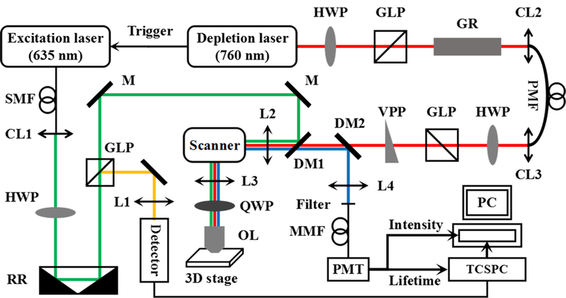
FIGURE 1.

Schematic diagram of the STED-FLIM imaging system. L, lens; CL, collimation lens; HWP, half wave plate; GLP, Glan-laser polarizer; GR, glass rod; RR, retro reflector; M, mirror; VPP, vortex phase plate; DM, dichroic mirror; OL, objective lens; QWP, quarter wave plate; SMF, single-mode fiber; PMF, polarization-maintaining fiber; MMF, multimode fiber; PMT, photomultiplier tube; STED, stimulated emission depletion
