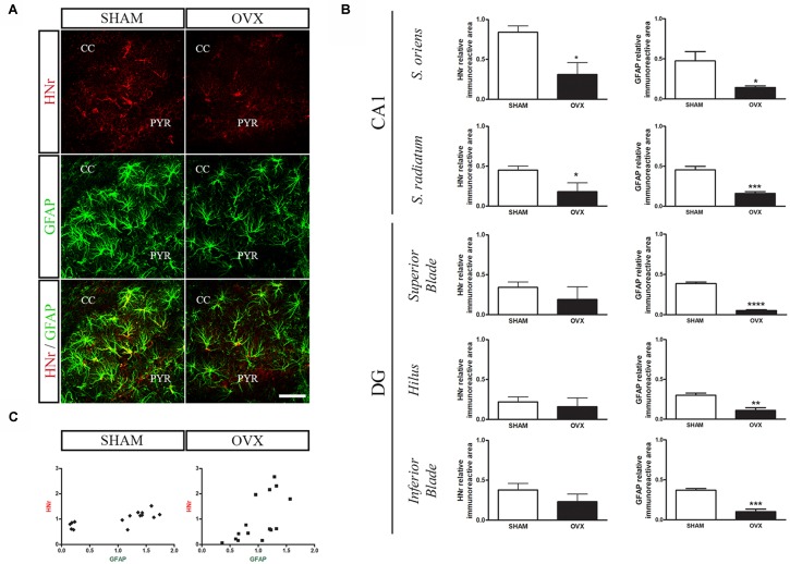
Figure 4.
Effects of long-term ovarian hormone deprivation on HNr and glial fibrillary acidic protein (GFAP) immunoreactivity in the hippocampus. Coronal sections from the hippocampus of SHAM and OVX animals were processed for double immunohistochemistry for HNr and GFAP. (A) Representative confocal microphotographs from CA1 stratum oriens (600×) show the expression of HNr (red), GFAP (green) and the merged image (yellow). (B) Quantification of relative immunoreactive area (immunoreactive area/total area) for HNr and GFAP using ImageJ software. Each column represents the mean ± SEM of relative immunoreactive area (n = 3 animals/group). *p < 0.05, **p < 0.01, ***p < 0.001, Student’s t-test. (C) HNr and GFAP expression levels expressed as relative immunopositive area for each protein were normalized with respect to each corresponding hippocampal subregion (SHAM r = 0.74, p < 0.01; OVX r = 0.63, p < 0.05, Pearson correlation test). PYR, pyramidal layer; CC, corpus callosum; DG, dentate gyrus. Scale bar = 50 μm.

