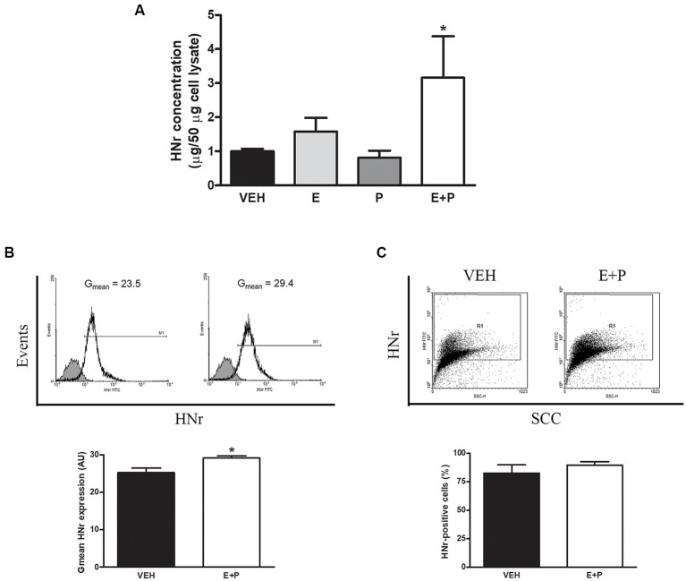
Figure 5.
HNr production and release by astrocytes in vitro. Cultured astrocytes were incubated with estradiol (E) and progesterone (P). HNr secreted levels were determined in conditioned media by ELISA. Harvested cells from additional cultures were immunostained for HNr and analyzed by flow cytometry. Each column represents the mean ± SEM of (A) the concentration of HNr in conditioned media normalized to 50 μg of total protein in corresponding cell lysate, (B) the fluorescence intensity of HNr staining (Gmean) or (C) the percentage of HNr-positive cells. The upper panels in (B,C) show representative histograms and dot plots of HNr expression in astrocytes incubated with VEH or E + P (n = 3–4 wells per group from three independent experiments). (A) *p < 0.05 vs. respective control without E; ANOVA followed by Tukey’s test, (B,C) *p < 0.05; Student’s t-test.

