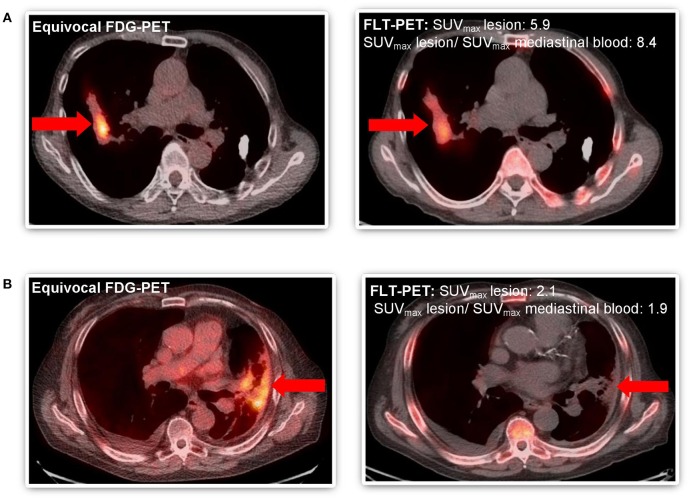
Figure 2.
Comparative imaging of FDG-PET and FLT-PET in patients with equivocal follow-up FDG-PET scan after SABR to thoracic malignancy. (A) Patient #4 WITH disease recurrence predicted by FLT-PET and confirmed by progression on subsequent serial imaging (both SUVmaxlesion >2.0 and ratio SUVmax lesion and mediastinal blood pool >2.0) and focal FLT uptake. (B) Patient #8 WITHOUT disease recurrence predicted by FLT-PET and confirmed by biopsy (SUVmaxlesion >2.0 but ratio SUVmax lesion and mediastinal blood pool < 2.0). Equivocal lesion by FDG-PET highlighted by arrow ( ).
).

