

Abstract
Aim:
To determine the degree of association between visual and tactile methods of caries removal compared with caries detector dye and laser fluorescence device (DIAGNOdent), which detects the degree of demineralization; to determine the presence of Streptococcus mutans via culture and polymerase chain reaction (PCR) techniques; and to find a suitable method for caries removal.
Materials and Methods:
A total of 75 patients were divided into three groups: visual and tactile (Group A), visual and tactile with caries detector dye (Group B), and visual and tactile with caries detector dye along with laser florescence readings (Group C). Caries removal was carried out using visual and tactile methods, caries detector dye, and laser fluorescence, and the samples obtained were subjected to culture and PCR. The data obtained were statistically analyzed using Pearson’s chi-square test, analysis of variance (ANOVA), and Tukey’s post hoc test.
Results:
Visual and tactile along with caries detector dye and laser florescence (Group C) is the most efficient method for caries removal.
Conclusion:
Caries detector dye along with visual, tactile examination and laser fluorescence is a valuable and superior tool for clinicians that aids in better caries removal and can prevent the overzealous removal of tooth structure.
KEYWORDS: Caries detector dye, caries removal, laser florescence (LF), MID, PCR, Streptococcus mutans
INTRODUCTION
During cavity preparation of a carious tooth for restoration, an attempt to excavate all the affected tissue in order to prevent secondary caries and to provide sound structural base for the future restoration assures removal of all caries-affected dentin, which has been conventionally assessed using a mouth mirror and an explorer. This subjective assessment has been shown to have limitations such as incomplete caries removal, which leads to secondary caries under the restorations and ultimate failure of treatment.[1] Caries detector dyes (CDD) have been developed to indicate areas of demineralization in order to aid in correct removal of infected dentin during cavity preparation. Laser fluorescence (LF) device DIAGNOdent (DD) is a non-invasive tool to detect demineralization zones in order to assess the carious state and the completeness of caries removal.[2] Polymerase chain reaction (PCR) detects even a small amount of bacteria and is a very sensitive method in which two primers mark the boundary of the gene for in vitro amplification. PCR can be used to detect Streptococcus mutans directly from the collected samples.[3] The aim of this in vivo study is to determine the degree of association between visual and tactile methods of caries removal compared with CDD and LF device. DIAGNOdent (DD) (KaVo Dental, Biberach, Germany) is a laser-based instrument used for the detection and quantification of dental caries. This instrument was calibrated according to the manufacturer’s specifications to determine the presence of Streptococcus mutans in the collected samples via culture and PCR techniques. This study focuses on the principle of minimally invasive dentistry (MID) and compares various methods of caries removal, thereby forming the basis of adhesive dentistry, in order to find a suitable method for caries removal with respect to the original tissues.
MATERIALS AND METHODS
This in vivo study includes 75 patients clinically examined for the presence of moderate dental caries (ICDAS 4). These patients were selected from those undergoing dental restorative treatment (for primary carious lesions) in Chettinad Dental College and Research Institute, Chennai, India. Institutional ethical committee clearance was obtained before the commencement of the study (IHEC/03/June2014/Desp.no.404-12.06.2014). Visual and tactile examination of the relevant dental carious lesion was done and radiographs were taken to assess the caries extension. On the basis of the results obtained, the patients were equally divided into three groups. A consent form was obtained from the patients prior to treatment. Molars and premolars were chosen in the study as they are more susceptible towards tooth decay as these teeth have lots of grooves, pits and fissures that can accumulate food particles.[4]
Group A: For 25 patients, caries removal was done with visual and tactile only and the samples were subjected to culture and PCR.
Group B: For 25 patients, caries removal was done with visual and tactile along with caries detector dye and the samples were subjected to culture and PCR.
Group C: For 25 patients, caries removal was done with visual and tactile and LF readings preoperatively, during, and postoperatively along with caries detector dye and the samples were subjected to culture and PCR.
Inclusion criteria
Moderate caries ICDAS 4 involved in posterior teeth
Moderate caries risk
Presence of at least one open carious lesion into dentin on occlusal surface
Age: 18–35 years
For Group C, LF readings preoperatively between 25 and 99
Exclusion criteria
Primary/mixed dentition
High caries risk
Teeth with restorations
Pulp involvement and periapical pathology
Tooth with development anomalies and disturbances
Systemic illness
Procedure
Group A: Visual and tactile examination: Under rubber dam isolation of the carious tooth, cavity preparation was done with the visual and tactile method using airotor, and final removal of carious dentin was carried out using a low-speed micromotor. Then, the cavity was assessed visually using a mouth mirror and probe/explorer to ensure satisfactory residual caries removal by both optical and tactile assessment, which includes texture (hard or soft), color (dark brown, light brown-black, dark yellowish, white), and appearance (shiny/dull). Dry/wet[5] caries removal was considered as complete when the explorer did not stick to the dentin and there was no tug-back sensation.[6] Dentin samples were then slowly taken using Rosehead bur no. 2 and subjected to culture and PCR.
Group B: Visual and tactile along with caries detector dye: Under rubber dam isolation of the carious tooth, cavity preparation was done with the visual and tactile method using airotor. At this stage, CDD (1% acid red with propylene glycol, Kuraray Co. Ltd., Japan) was applied to detect any infected dentin. Demineralized tissue appears as areas of pink purple staining.[5,7] The collected dentin samples were subjected to culture and PCR.
Group C: Visual and tactile along with caries detector dye and laser fluorescence: Under rubber dam isolation, the first LF readings were taken prior to cavity preparation. After conventional cavity preparation, the second LF readings were taken. At this stage, CDD (1% acid red with propylene glycol, Kuraray Co. Ltd., Japan) was applied to detect any infected dentin. Demineralized tissue appears as areas of pink purple staining.[5,6] Again, after removal of dye-stained tissue, the third LF readings were taken and tabulated. The collected dentin samples were subjected to culture and PCR. DIAGNOdent value interpretation given by Lussi et al.[8] was considered as the guideline.
Collection of samples
Standardized samples were obtained based on the methods of Kidd et al.[4] from each site in all the three groups. Dentin samples were taken from the sites and transferred to Eppendorf tubes containing Brain Heart Infusion Agar and 0.2 units/mL of Bacitracin.[7] The extracted dentin samples were subjected to culture and PCR. The presence of S. mutans was confirmed on the basis of characteristic colony, morphology, and standard biochemical tests.[4] The culture grown was compared morphologically with standard reference strain SS909 obtained from Department of Microbiology, CMC, Vellore (freeze-dried and lyophilized) [Figure 1].
Figure 1.
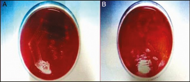
(A) Standard culture and (B) cultures from clinical samples
DNA extraction procedure
Chromosomal DNA was extracted from samples by high salt method,[9] and the extracted DNA was subjected to amplification and investigated by agarose gel electrophoresis. DNA Amplification Kit (GeNei, Bengaluru) was used for DNA amplification. Primers for this study for PCR were selected on the basis of spaP gene from S. mutans strain NG5.[3]
Agarose gel electrophoresis
The PCR product was analyzed electrophoretically for the presence of DNA, which can be seen under UV light that fluoresces as bands and photographed under UV light using a Polaroid camera (Fotodyne, Hartland, Wisconsin, USA). PCR was used to amplify a sequence (192 bp) of the spaP gene, which encodes surface protein antigen I/II of S. mutans[10] [Figure 2]. Statistical analysis of the results obtained from the clinical samples after culture and PCR amplification was conducted using the IBM SPSS software (version 21.0). Pearson’s chi-square test, ANOVA, and Tukey’s post hoc test (Tukey’s HSD test) were also carried out in the present study.
Figure 2.

(A) Polymerase chain reaction amplification of spaP gene sequence (192BP) from standard culture (B) Negative from clinical samples (C) Positive clinical samples
RESULTS
Pearson’s chi-square test [Table 1] was conducted to compare the PCR and culture values in all the three groups, and the results were found to be statistically significant (P = 0.000). Inter-group comparison of PCR and culture values showed a significant drop in Group C.
Table 1.
Comparison of polymerase chain reaction and culture in all groups (chi-square test)
| Group | Growth | PCR | Culture | P value |
|---|---|---|---|---|
| 1 | + | 12 | 10 | 0.000 |
| − | 13 | 15 | ||
| 2 | + | 12 | 6 | 0.000 |
| − | 13 | 19 | ||
| 3 | + | 7 | 7 | 0.000 |
| − | 18 | 18 |
ANOVA [Table 2] was carried out to compare the LF readings taken preoperatively, during, and postoperatively after complete caries removal, and the corresponding mean values were 40.96, 19.80, and 11.04. Statistical significance was set at P = 0.000.
Table 2.
Comparison of laser fluorescence values between intervals (analysis of variance test)
| Group | N | Mean | Standard Deviation | P value |
|---|---|---|---|---|
| Pre | 25 | 40.96 | 14.653 | |
| During | 25 | 19.80 | 4.444 | 0.000 |
| Post | 25 | 11.04 | 3.372 | |
| Total | 75 | 23.93 | 15.479 |
Tukey’s HSD test (post hoc) test was conducted to achieve multiple comparisons between LF readings preoperatively, during, and postoperatively, and the result was found to be statistically significant (P = 0.000) [Table 3].
Table 3.
Tukey HSD test (post hoc) multiple comparison between preoperative, during, and postoperative laser fluorescence readings
| Group | Comparison | Mean difference | P value |
|---|---|---|---|
| Pre | During | 21.160 | 0.000 |
| Post | 29.920 | 0.000 | |
| During | Pre | −21.160 | 0.000 |
| Post | 8.760 | 0.003 | |
| Post | Pre | −29.920 | 0.000 |
| During | −8.760 | 0.003 |
DISCUSSION
Worldwide, it is estimated that 50–70% of a general dentist’s activities lies in the replacement of existing restorations.[11] On the other hand, use of CCD and its staining may lead to overzealous tooth structure removal.[12] Caries removal method should be chosen as it preserves the natural tissues of patients, thus following the principles of MID. All clinical stages of restorative procedures should be optimized, with prevention of disease recurrence as the ultimate goal of modern dentistry.[13,14] In this study, we have used LF device DD to assess the carious state and the completeness of caries removal, as it is non-invasive, sensitive, and reproducible between examiners[8] The PCR method provides a more specific, more sensitive, and simpler means of detecting cariogenic bacterial species compared to other conventional techniques.[15] According to de Souza et al.,[16] LF pen has higher accuracy in detecting both enamel and dentin carious lesions than bite wing radiographs. Goswami and Rajwar[17] showed that DD is a reproducible device that is useful for secondary caries detection in posterior teeth as an adjunct to visual examination. Castilho et al.[18] showed that the ability of DD to detect initial carious lesions was higher than that of ICDAS, but with low specificity. In previous in vitro studies, which have evaluated the performance of LF for residual caries detection in primary teeth after different caries removal methods, it has been reported that the LF system could be effective in assessing caries removal by different methods.[19,20] In our study, Group A showed [Table 1] 12 PCR positive and 10 culture positive (48% and 40%, respectively), Group B showed 12 PCR positive and 6 culture positive (48% and 24%, respectively), and Group C showed 7 PCR positive and 7 culture positive (28% and 28%, respectively).
The comparison between the groups is significant (P = 0.000). Visual and tactile Group A has a large number of positive for PCR and culture; CDD Group B has equal number of positive detection in PCR and less number in culture; and Group C has the least number of positive detection in PCR and culture. This clearly shows the efficacies of the different methods. In our study, of the 75 patients, Groups B and C, where there was no staining, were subsequently inferred by culture test and PCR as negative. It means that areas unstained by the dye are 100% free of infected layer. In the remaining patients, staining by the dye can be considered as positive response and subjected to culture and PCR. The results showed that 13 patients were culture positive and 19 were PCR positive.
A total of 25 patients (Group C) were subjected to DD observation on an experimental basis, of whom 8 fell in the category of no staining by CDD. The LF readings range was 8–16 or less, and the culture and PCR were also negative. In the absence of dye staining, negative culture, and PCR, visual and tactile method of caries removal is found to produce satisfactory LF readings in the range of 8–16. In the cases where dye has stained, five patient samples showed culture and PCR negative LF readings in the range of 17–19. For any reading below 19, even when dye is able to stain, both PCR and culture were negative. In the remaining 12 patients where LF was used, both culture and PCR were positive along with staining with CDD and LF value ranging from 20 to 36, which requires carious tissue removal. These readings along with culture and PCR findings represent the best evidence currently available; they should not be considered as rigid threshold, rather they should help to guide make a clinical decision whether to cut stained dentin or not. Hence, LF reading above 20 warrants the use of caries detector dye and removal of stained dentin. From the inference of the current study, when the LF reading goes below 20, there is no need to use a CDD. When the DD readings are above 20 (second readings), it is necessary to use CDD and remove all stained areas, and the samples become culture and PCR positive. Hence, removing the stained dentin in these patients is justified to prevent secondary caries. Staining by CDD need not necessarily mean the presence of S. mutans, since PCR was positive only in 40% of cases stained by the CDD. In the absence of dye staining, visual and tactile methods of caries removal can be considered satisfactory. The results clearly show the degree of association between visual and tactile criteria and LF readings. Ntovas et al.[21] in a recent study concluded that the participants’ ability to diagnose residual caries during cavity preparation was independent of their experience. During the removal of carious lesions, with the goal of limiting the sacrifice of healthy structures and the leftover of residual caries, clinicians should combine a selective removal technique and the attentive visual inspection with at least one further diagnostic method, aside from the dental explorer. LF readings along with clinical experience lead to judicious removal of dentin, thus following the principles of MID.
CONCLUSION
The results of our study provide a clear degree of association between visual and tactile methods and LF readings. Hence, we conclude that because of its non invasive nature leading to successful restoration, utilization of LF along with clinicians experience aids in judicious caries removal and preventing overzealous removal of dentin, thus protecting natural tissues.
Financial support and sponsorship
Nil.
Conflicts of interest
There are no conflicts of interest.
REFERENCES
- 1.Anderson MH, Charbeneau GT. A comparison of digital and optical criteria for detecting carious dentin. J Prosthet Dent. 1985;53:643–6. doi: 10.1016/0022-3913(85)90011-3. [DOI] [PubMed] [Google Scholar]
- 2.Lussi A, Imwinkelried S, Pitts N, Longbottom C, Reich E. Performance and reproducibility of a laser fluorescence system for detection of occlusal caries in vitro. Caries Res. 1999;33:261–6. doi: 10.1159/000016527. [DOI] [PubMed] [Google Scholar]
- 3.Ono T, Hirota K, Nemoto K, Fernandez EJ, Ota F, Fukui K. Detection of Streptococcus mutans by PCR amplification of spaP gene. J Med Microbiol. 1994;41:231–5. doi: 10.1099/00222615-41-4-231. [DOI] [PubMed] [Google Scholar]
- 4.Yadav K. Dental caries: A microbiological approach. J Clin Infect Dis Pract. 2017;2:118. [Google Scholar]
- 5.Kidd EA, Joyston-Bechal S, Beighton D. Microbiological validation of assessments of caries activity during cavity preparation. Caries Res. 1993;27:402–8. doi: 10.1159/000261571. [DOI] [PubMed] [Google Scholar]
- 6.Bohari MR, Chunawalla YK, Ahmed BM. Clinical evaluation of caries removal in primary teeth using conventional, chemomechanical and laser technique: An in vivo study. J Contemp Dent Pract. 2012;13:40–7. doi: 10.5005/jp-journals-10024-1093. [DOI] [PubMed] [Google Scholar]
- 7.Kidd EA, Joyston-Bechal S, Beighton D. The use of a caries detector dye during cavity preparation: A microbiological assessment. Br Dent J. 1993;174:245–8. doi: 10.1038/sj.bdj.4808142. [DOI] [PubMed] [Google Scholar]
- 8.Lussi A, Hibst R, Paulus R. DIAGNOdent: An optical method for caries detection. J Dent Res. 2004;83:C80. doi: 10.1177/154405910408301s16. [DOI] [PubMed] [Google Scholar]
- 9.Kidd EA, Joyston-Bechal S, Smith MM, Allan R, Howe L, Smith SR. The use of a caries detector dye in cavity preparation. Br Dent J. 1989;167:132–4. doi: 10.1038/sj.bdj.4806939. [DOI] [PubMed] [Google Scholar]
- 10.Allaker RP, Seddon SV, Tredwin C, Lynch E. Detection of Streptococcus mutans by PCR amplification of the spaP gene in teeth rendered caries free. J Dent. 1998;26:443–5. doi: 10.1016/s0300-5712(97)00058-4. [DOI] [PubMed] [Google Scholar]
- 11.Gold OG, Jordan HV, Van Houte J. A selective medium for streptococcus mutans. Arch Oral Biol. 1973;18:1357–64. doi: 10.1016/0003-9969(73)90109-x. [DOI] [PubMed] [Google Scholar]
- 12.Yip HK, Stevenson AG, Beeley JA. The specificity of caries detector dyes in cavity preparation. Br Dent J. 1994;176:417–21. doi: 10.1038/sj.bdj.4808470. [DOI] [PubMed] [Google Scholar]
- 13.Ericson D. The concept of minimally invasive dentistry. Dent Update. 2007;34:9–10. doi: 10.12968/denu.2007.34.1.9. 12-4, 17-8. [DOI] [PubMed] [Google Scholar]
- 14.Tyas MJ, Anusavice KJ, Frencken JE, Mount GJ. Minimal intervention dentistry—a review. FDI commission project 1-97. Int Dent J. 2000;50:1–12. doi: 10.1111/j.1875-595x.2000.tb00540.x. [DOI] [PubMed] [Google Scholar]
- 15.Hongkun W, Mingwen F, Xuendong Z, Zhuan bian AM, Zhi chen QZ. Detection of Streptococcus mutans and Streptococcus sobrinus on the permanent first molars of the Mosuo people in China. Caries Res. 2003;37:374–80. doi: 10.1159/000072171. [DOI] [PubMed] [Google Scholar]
- 16.de Souza JF, Diniz MB, Boldieri T, Rodrigues JA, Lussi A, de Cássia Loiola Cordeiro R. In vitro performance of a pen-type laser fluorescence device and bitewing radiographs for approximal caries detection in permanent and primary teeth. Indian J Dent Res. 2014;25:702–10. doi: 10.4103/0970-9290.152165. [DOI] [PubMed] [Google Scholar]
- 17.Goswami M, Rajwar AS. Evaluation of cavitated and non-cavitated carious lesions using the WHO basic methods, ICDAS-II and laser fluorescence measurements. J Indian Soc Pedod Prev Dent. 2015;33:10–4. doi: 10.4103/0970-4388.148961. [DOI] [PubMed] [Google Scholar]
- 18.Castilho IS, Fymd C, Bueno AC, Moreiro AN, Ferreira EF, Magalh-Aes CS. Valiadation of DIAGNOdent laser fluorescence and the international caries detection and assessment system (ICDAS) in diagnosis of occlusal caries in permanent teeth: An vitro study. Eur J Oral Sci. 2016;124:188–94. doi: 10.1111/eos.12257. [DOI] [PubMed] [Google Scholar]
- 19.Almaz ME, Sönmez IŞ, Oba AA. Comparison of chemomechanical caries removal using Papacárie versus conventional method in children. Eur J Gen Dent. 2016;5:1–5. [Google Scholar]
- 20.Spaveras A, Tsakanikou A, Karkazi F, Antoniadou M. Caries detection with laser fluorescence devices. Limitations of their use. Stoma Edu J. 2017;4:46–53. [Google Scholar]
- 21.Ntovas P, Loubrinis N, Maniatakos P, Rahiotis C. Evaluation of dental explorer and visual inspection for the detection of residual caries among Greek dentists. J Conserv Dent. 2018;21:311–8. doi: 10.4103/JCD.JCD_67_17. [DOI] [PMC free article] [PubMed] [Google Scholar]